Research Article | | Peer-Reviewed

Synthesis, In vitro Antimicrobial, and In Silico Studies of 2-Hydroxy-1, 2-Diphenylethanone Derivatives

Received: 7 August 2026     Accepted: 19 August 2026     Published: 9 September 2026
Views:       Downloads:
Abstract

Infectious diseases place a significant impact on the healthcare system. It causes 7.7 million fatalities each year, with a disproportionately high burden in sub-Saharan Africa. Drug resistant pathogens attributed to 4.95 million and 1.27 million are illnesses of bacteria that are resistant to the current medications. Therefore, there is an urgent need for new, safe, and effective compounds to combat antimicrobial resistance. The aim of this study was to synthesize new compounds derived from 2-hydroxy-1, 2-diphenylethanone using the Mannich reaction and evaluate their antimicrobial activities against 26 bacterial strains and 4 fungal strains. Three compounds were successfully synthesized and their structures were confirmed as 3 (diethylamino)-2-hydroxy-1, 2-diphenylpropan-1-one (2) 1-(2-hydroxy-3-oxo-2, 3 diphenylpropyl) urea (3), and 2-hydroxy-1, 2-diphenyl-3-(piperidin-1-yl) propan-1-one (4), using  1H and 13C-NMR spectroscopy. All the synthesized compounds exhibited broad-spectrum antibacterial activity. Compound (3) demonstrated the highest activity, with MIC of 10 µg/mL against Shigella sonnei 1, Shigella boydii D13629, and Pseudomonas aeruginosa MDR1 and compound (4) demonstrated the highest antifungal activity, with MIC of 200 µg/mL against Candida albicans ATCC 10231, Aspergillus niger ATCC 6275, Penicillium funiculosum NCTC 287, and Penicillium notatum ATCC 11625. Molecular docking showed the compounds interact favorably with conserved residues in the binding site of E. coli DsbA (PDB ID: 8DN0) through hydrogen bonding and hydrophobic interactions, with docking scores of –7.5 kcal/mol compound (3), –7.1 kcal/mol compound (4), and –6.5kcal/mol compound (2). This research suggests verify the molecular docking results experimentally and biological activities, such as antiviral, anticancer, anti-inflammatory, and antioxidant properties and evaluate the safety profile of the produced chemicals.

Published in World Journal of Health Services Research (Volume 1, Issue 1)
DOI 10.11648/j.wjhsr.20260101.11
Page(s) 1-13
Creative Commons

This is an Open Access article, distributed under the terms of the Creative Commons Attribution 4.0 International License (http://creativecommons.org/licenses/by/4.0/), which permits unrestricted use, distribution and reproduction in any medium or format, provided the original work is properly cited.

Copyright

Copyright © The Author(s), 2026. Published by Science Publishing Group

Keywords

2-hydroxy-1, 2-diphenylethanone, Antimicrobial Activity, In-silico Studies, Disulfide Bond Oxidoreductase, Mannich Reaction

1. Introduction
Infectious diseases are caused by microorganisms such as bacteria, viruses, fungi or parasites and continue to exert a substantial strain on healthcare systems . It places a significant strain on the healthcare system. In 2022, infectious diseases affected hundreds of millions of people and accounted for an estimated 7.7 million deaths worldwide, with a particularly high burden in Sub-Saharan Africa. Bacterial infections range from asymptomatic to life threatening disease. .
Benzoin (2-hydroxy-1, 2-diphenylethanone) (1) is organic molecule with ketone, alcohol, and aromatic phenyl functional groups. It possesses several pharmacological properties, including antimalarial, antifungal, antioxidant, anticancer, and antibacterial properties . As a natural product, benzoin is obtained from the resin of Styrax species and has long been used in perfumery, incense and traditional medicine for wounds, skin conditions and a variety of ailments .
Derivatives of benzoin have been prepared by various methods (for example, Schiff base formation) and have shown antimicrobial promise; examples include N-(2-hydroxybenzylidene)-2-hydroxyimines and sulfamethazine derivatives . These findings have stimulated interest in benzoin scaffolds for the synthesis of biologically active compounds .
Antimicrobial resistance (AMR) is a growing global health threat with increasing human and economic costs . The shortage of newly approved antibiotics and the emergence of novel resistance mechanisms underscore the need for new chemical entities and continued research and development .
Disulfide bond formation is an essential post-translational modification for many secreted bacterial proteins. DsbA proteins are thiol–disulfide oxidoreductases that catalyse intrachain disulfide bond formation and are present in diverse bacteria, including Klebsiella pneumoniae, Vibrio cholerae, Pseudomonas aeruginosa, Staphylococcus aureus, Neisseria meningitidis, Bacillus subtilis, Wolbachia pipientis and Burkholderia pseudomallei . The aim of this work was to synthesise derivatives of 2-hydroxy-1, 2-diphenylethanone and evaluate their antimicrobial potential using in vitro assays and in silico molecular modelling.
2. Methods
2.1. Chemical Material and Apparatus
All solvents (methanol, n-hexane, chloroform, diethyl ether, ethanol) were HPLC grade and obtained from Loba Chemie. Other reagents (benzoin, 2, 4-dinitrophenylhydrazine, urea, formaldehyde, piperidine, N, N-diethylamine, etc.) were obtained from the Addis Ababa University School of Pharmacy laboratory. Thin-layer chromatography (TLC) was performed on pre-coated silica plates (POLYGRAM SIL G/UV254, 0.2 mm layer). Instrumentation included a rotary evaporator (Heidolph Laborota 4001), vacuum pump (BUCHI), UV-visualiser (CAMAG), electronic balance (Sicentec), incubator, autoclave and biosafety cabinet. NMR spectra were recorded on a Bruker Avance 400 MHz (1H) and 100 MHz (13C) spectrometer, TMS was used as internal standard and CDCl3 as solvent.
2.2. Synthesis
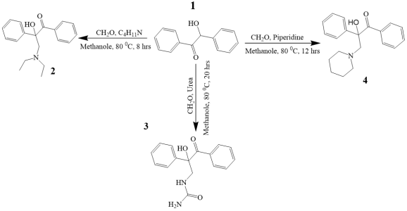
General synthesis of 2-hydroxy-1, 2-diphenylethanone derivatives was achieved using Mannich type conditions starting from benzoin. . General scheme and conditions see Figure 1 below.)
Scheme 1. General synthesis of 2-hydroxy-1, 2-diphenylethanone derivatives.
2.2.1. Synthesis of 3-(Diethyl Amino)-2-Hydroxy-1, 2 Diphenylpropan-1-One) (2)
Initially 30 mL of methanol to was added a 100 mL of reaction flask, then 0.83g (4.00 mmol) of Benzoin was added to the reaction flask containing methanol and stirred at 50°C by magnetic sterrier until it completely dissolves then added four drops of concentrated Sulfuric acid as catalyst. In another beaker, 0.36g (1.334mmol) of diethyl amino was dissolved in 20 mL methanol, and the solution was added stepwise to the reaction flask containing Benzoine. Then it was stirred at a temperature of 80°C at a rate of 500 revolutions per minute (rpm) and the reaction was monitored with TLC every 60 minutes for 8 hrs. Then, the product was poured in a round bottom flask and the solvent was removed by rotavouper at a temperature of 80°C and a rotation of 100 rpm until it dried. Then after, the dried product was collected and run a flash column chromatogram for further purification with an increasing gradient of hexane in ethylate as the eluting solvent.
2.2.2. Synthesis of 1-(2-Hydroxy-3-Oxo-2, 3-Diphenylpropyl) Urea (3)
At the beginning, 20 mL of methanol was added in to a 100 mL of reaction flask, then 0.85 mL of a 37% solution of formaldehyde then added four drops of concentrated sulfuric acid as catalyst. Then 0.6g of urea was added to the reaction flask. In another beaker 2.0 g/4.00 mmol of Benzoin was dissolved in 30 ml of methanol and added to the reaction flask and stirred at 50°C then the reaction was monitored with TLC every 60 minutes for 20-hrs. Then, the product was poured in a round bottom flask, and the solvent was removed by rotavouper at a temperature of 50°C and rotation of 100 revolutions per minute (rpm) until it dried. Then after, the dried product was collected and run a flash column chromatogram for further purification.
2.2.3. Synthesized of 2-Hydroxy-1, 2-Diphenyl-3-(Piperidin-1-yl) Propan-1-One (4)
Initially added 20 ml of methanol into a 100 mL of reaction flask, then 0.68 ml of a 37% solution of formaldehyde, and added four drops of concentrated Sulfuric acid as catalyst. Then 0.85g (2 ml) of Piperidine was added to the reaction flask. In another beaker 2.0 g/4.00 mmol of Benzoin was dissolved in 30 ml of methanol and added to the reaction flask and stirred at 80°C and the reaction was monitored with TLC every 60 minutes for 12 hrs. Then, the product was poured in a round bottom flask, and the solvent was removed by rotavouper at a temperature of 80°C and rotation of 100 revolutions per minute (rpm) until it dried. Then after, the dried product was collected and run a flash column chromatogram for further purification with an increasing gradient of hexane in ethylate as the eluting solvent.
2.3. Structural Confirmation of the Synthesized Compounds
The structures of compounds 2, 3 and 4 were identified by analyzing their 1H and 13C-NMR spectra. The chemical shifts are denoted in ppm and coupling constants (J) are expressed in Hz. Signal multiplicities are indicated as follows: s = singlet; d = doublet; t = triplet; ddd = doublet of doublets; and m = multiplet (Annex-II).
2.4. In Vitro Antimicrobial Assay.
2.4.1. Disk Diffusion Method
The disc diffusion method was used to screen the in vitro antibacterial assay by evaluating the zones of inhibition generated by the test samples. . The Disk Diffusion antimicrobial experiment was conducted according to the CLSI guideline. All culture media used were prepared according to the manufacturer instruction. The media was sterilized at 121°C for 15 minutes using an autoclave and allowed cool to 45°C 50°C in water bath. Then 20 ml of media was poured into 100 mm diameter size petri-dishes under aseptic condition inside level II Bio-safety Cabinet and allowed some time to solidify. For antibacterial experiments 4-5 bacterial colony of each tested organisms were inoculated with inoculating loop in to Petri dishes containing agar medium. Each bacterial test organisms were incubated for 18-24 h at 37°C. Standardization was accomplished by preparing a suspension of 3-5 inoculations from a fresh, pure culture of the specimen bacterium in Mueller Hinton broth. The produced suspension's absorbance was measured using an ultraviolet-visible (UV) spectrophotometer at 625 nm having a path length of 1cm until the absorbance reading was 0.08 to 0.1, which is proportional to 1x10⁸ CFU/ml bacteria. The standardized solution was then further diluted with Muller Hinton Broth at 1: 100 ratios to yield a colony suspension containing 1x10⁶ CFU/mL bacteria. For the broth dilutions, final suspensions of 5 x10⁵ CFU/mL were employed, and evaluate the antibacterial activity of the compounds in next broth dilution experiment .
By making a stock solution of 1 mg/mL of the benzoin derivatives in 1% DMSO, the antibacterial and antifungal effects of the chemicals produced were investigated. The antibacterial and antifungal activity test values ranged from 5 to 800μg/mL for antibacterial and from 50 to 2000 μg/mL for antifungal. Then the stock solution was diluted in the proper amounts of distilled water. After that, sterile Petri dishes were filled with molten media and incubated for 24 hrs at 37°C to check for contamination in order to create the serial nutritional agar plates. After serial nutrient production, the inoculums were evenly swabbed and left to dry for five minutes. The Petri plates were subsequently incubated for 24 hrs at 37°C, and the diameter of the inhibitory zone was determined in millimeters. The experiment was performed using 1% DMSO as a negative control and ciprofloxacin as a positive control for bacteria. The Zone of inhibition (ZOI) was then compared.
On Saborauds dextrose media, the test samples' antifungal potential was evaluated using an identical methodology against the fungal infections. Millimeters were used to measure the ZOI's diameter in the Petri plates following three days of room temperature incubation. The Griseofulvin was used in this instance as the standard drug.
2.4.2. Broth Dilution Method
The minimum inhibitory concentration (MIC) of the produced compounds was determined using the broth dilution procedure .
A 100μl of Muller Hinton broth was added into each well of the 96-well microplates. From a stock solution, the serial dilutions were made ranging from 800 µg/ mL to 5 µg/mL using Mueller–Hinton broth in 96-well microplates. Hence concentrations of 5, 10, 25, 50, 100, 200, 400 and 800 µg/ mL of synthesized compounds were made for antibacterial activity test. The bacterial suspension containing approximately 5 × 10⁵ CFU/ml was prepared from a refreshed culture. From this suspension, 100 μl was inoculated into each well and incubated for 18-24 hour at 37°C. After incubation, the MIC values were visually determined by observing the presence and absence of turbidity. Absence of turbidity in each well marked the MIC. A negative control of 1% DMSO was used for the experiment.
A sterility control, which involves growth control using nutritious broth and DMSO devoid of antibiotics, was also carried out. Each test well and growth control well was cultured for three days at 25°C for fungi and for twenty-four hours at 37°C for bacteria. MIC was indicated by the test tubes' lack of turbidity .
2.5. In Silica Study
2.5.1. Molecular Docking
A protein-ligand docking technique called CB-Dock automatically finds the binding sites, determines their size and center, and adjusts the docking box's dimensions based on the query ligands. The hit percentage and accuracy of blind docking can be improved by cavity-focused docking, according to extensive benchmarking. As a result, CB-Dock can speed up the docking process and increase accuracy by employing our curvature-based cavity detection method to forecast the target proteins' binding locations . CB-Dock workflow comprised six stages: Input and Preprocessing, Cavity Detection, Grid Generation, Docking Simulation with Autodock Vina, Score Ranking & Results Visualization and Output. For validation of the result redocking was performed for both native ligand and the synthesized compounds.
2.5.2. Pharmacokinetics and Drug-likeness Properties
With the aid of the online software tool ADMET lab 3.0, the physicochemical, pharmacokinetic, and toxicity characteristics of the produced compounds were anticipated, as described by J. Dong et al 2013 .
3. Result and Discussion
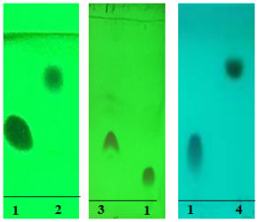
In this study, three compounds, 3-(diethylamino)-2-hydroxy-1, 2-diphenylpropan-1-one (2), 1-(2-hydroxy-3-oxo-2, 3-diphenylpropyl)urea (3), and 2-hydroxy-1, 2-diphenyl-3-(piperidin-1-yl)propan-1-one (4), were successfully synthesized from 2-hydroxy-1, 2-diphenylethanone using a one-pot reaction. The synthesis followed a Mannich-type reaction pathway, leading to the formation of β-amino alkylating products. The physical properties of these compounds are shown in Table 1: below. The Rf value for the three of synthesized compounds as indicated in Table 1 blew, you can see their pictures in Figure 3 Annex-I.
Table 1. Physical properties of the synthesized compounds.

Compound

MF

MW

Color

PS

Yield

Rf value

2

C19H23NO2

297.37

White

Crystal

56%

0.7

3

C17H17NO3

284.31

White

Crystal

24%

0.4

4

C20H23NO2

309.40

Yellowish

Powder

64%

0.8

Key: MF=Molecular formula, MW= Molecular weight, PS= Physical state, Rf = Retention factor
3.1. Structural Confirmation of 3-(Diethylamino)-2-Hydroxy-1, 2-Diphenylpropan-1-One (2)
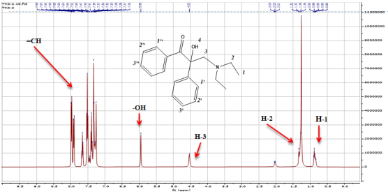
3-(Diethylamino)-2-hydroxy-1, 2-diphenylpropan-1-one was successfully synthesized as a white crystal (56% w/w M.wt 297.37) with an Rf value of 0.7 The 1H-NMR spectrum displayed a triplet at δ 1.24 ppm (3H, J=6.7 Hz, H-1) and a quartet at δ 2.96 ppm (2H, J=6.8 Hz, H-2), consistent with an ethyl group (–CH₂CH₃) moiety. A pair of doublets at δ 3.00 and 3.08 ppm (H-3) suggested diastereotopic methylene protons adjacent to an electronegative group or stereocenter. A singlet at δ 6.00 ppm corresponded to a hydroxyl (OH) proton, indicating the presence of a free phenolic or alcoholic group. Aromatic proton signals appeared as multiplets and doublets in the region δ 7.40–7.94 ppm, characteristic of substituted aromatic rings (See Figure 1 below).
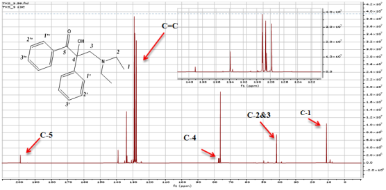
The 13C-NMR (126 MHz, CDCl3) demonstrates the presence of two methyl groups C-1 (𝛿=11.01), the presence of methylene carbon C-2&3 ((𝛿=41.73 & 50.93), also quaternary carbon at (𝛿=77.44) and aromatic carbons (See Figure 2). Thus, the structure of compound (2) was confirmed by both 1H-NMR and 13C-NMR investigations as 3-(diethylamino)-2-hydroxy-1, 2-diphenylpropan-1-one.
Figure 1. 1H-NMR data for the synthesized compound (2).
Key: =CH protons of aromatic rings, -OH proton of hydroxyl group on the quaternary carbon, H-3 methylene proton, H-2 Methylene proton, Methyl proton.
Key: C-5 Carbon of carbonyl Carbon, C=C aromatic carbon, C-4 quaternary Carbon, C-2&3 Methylene carbon, and C-1 Methyl Carbone.

Download: Download full-size image

Figure 2. 13C-NMR data for the synthesized compound (2).
3.2. Structural Confirmation of 1-(2-Hydroxy-3-Oxo-2, 3-Diphenylpropyl) Urea (3)
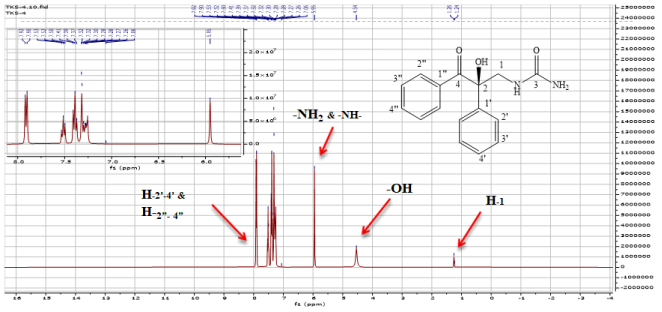
1-(2-Hydroxy-3-oxo-2, 3-diphenylpropyl) urea was successfully synthesized as a white crystal (24% w/w, M. wt 284.31) with an Rf value of 0.4 on TLC using HE/CF (2: 1) as the mobile phase. The 1H NMR (500 MHz, CDCl3) of (3) shows a total of ten sets of protons. Among those, one methylene proton assigned to H-1 (𝛿=4.45, d, J= =11.04Hz), three amine protons assigned to NH& &NH2 (𝛿=5.96, s,,) two methylene protons assigned to H-3 (𝛿=2.96, d,= J =11.9, Hz) one hydroxyl proton –OH (𝛿=6.00, and, s) sets set of aromatic protons on two phenyl rings for more information you can see Figure 4 (Annex –II).
The 13C-NMR (400 MHz, CDCl3) demonstrates the presence of one methylene carbon assigned to C-1 (𝛿=55.23), the presence one of quaternary carbon C-2 at (𝛿=77.23), carbonyl carbon of the amide at (𝛿=167.32), carbonyl carbon of the ketone at (𝛿=197.78) and aromatic carbons. Thus, the structure of compound (3) was confirmed by both 1H-NMR and 13C-NMR investigation, for more information see Figure 5 (Annex –II). Based on this information of both 1H-NMR and 13C-NMR structure of compound (3) was confirmed as 1-(2-Hydroxy-3-oxo-2, 3-diphenylpropyl) urea.
3.3. Structural Confirmation of 2-Hydroxy-1, 2-Diphenyl-3-(Piperidin-1-yl) Propan-1-One (4)
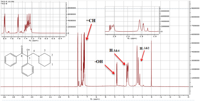
2-Hydroxy-1, 2-diphenyl-3-(piperidin-1-yl) propan-1-one was synthesized as a yellowish powder (64% w/w, M. wt 309) with an Rf value of 0.8 on TLC using HE/CF (2: 1) as the mobile phase. The 1H NMR (400 MHz, CDCl3) of (4) shows a total of eleven sets of protons. Among those eight methylene protons assigned to H1-4, one hydroxyl proton, ten aromatic protons on two phenyl rings. The two methylene protons assigned to H1 & 2 (𝛿 = 1.13 - 159, m, J = 6.7,) one hydroxyl proton H-4 (𝛿=3.74, s) for more information see Figure 6 (Annex – II)
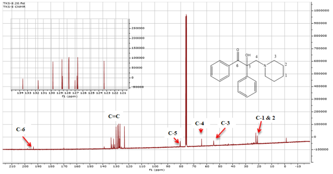
The 13C-NMR (101 MHz, CDCl3demonstratees the presences of four methylene groups C-1 (𝛿=11.01), the presence of methylene carbon & -2&3 ((𝛿 = 41.73 & 50.93), the quaternary carbon at (𝛿=7, 7.44) and aromatic carbons for more information see Figure 7 (Annex – II). Thus, the structure of compound (4) was confirmed by both 1H-NMR and 13C-NMR investigations as 2-hydroxy-1, 2-diphenyl-3-(piperidin-1-yl) propan-1-one.
3.4. Antimicrobial Activity of the Synthesized Compound
3.4.1. Antibacterial Activity
In this research, 2-hydroxy-1, 2-diphenylethanone derivatives were synthesized (2), (3), and (4) tested in vitro against 26 bacterial strains by both disc diffusion and broth dilution methods. Surprisingly, three of them were demonstrating similar antibacterial activity with MICs ranging from 10µg/mL to 800µg/mL and a zone of inhibition ranging from 6.0mm to 15.8mm. Among the three synthesized compounds, the maximum activity against Shigella soneii 1, Shigella boydii D13629, and Pseudomonas aeruginosa MDR1 which is 10µg/mL by (3) (See Table 2). The possible reason for this structural variations influence antimicrobial activity may be polar substituents maximum activity as well as good docking score compound (2) and (4).
Table 2. Anti-bacterial activity of synthesized compounds against the bacterial strain.

Bacteria strain

ZOI in mm (200µg/ml)

MIC (µg/ml)

2

3

4

Ciprofloxacin

2

3

4

E.coli K99

13.5±0.5

12.2±0.6

13.5±0.5

16.0±1.0

25

100

25

E.coli K88

14.8 ± 0.8

12.5±0.0

14.8±0.8

17.0 ±0.0

25

100

25

E.coli 306

14.0 ± 0.9

13.0±0.5

14.3±0.6

17.0 ± 1.0

25

100

25

E.coli LT37

14.0 ± 0.0

12.5±0.0

14.0±0.0

16.0 ± 0.0

25

100

25

E.coli 872

14.5 ± 0.5

12.7±0.3

14.5±0.5

15.8± 0.8

25

100

25

E.coli ROW 7/12

15.0±0.0

12.0±1.0

14.5±0.5

16.5 ±0.0

25

100

25

E.coli 3: 37C

15.2 ±0.3

11.5±0.5

15.2±0.3

16.5 ± 1.0

25

100

25

E.coli CD/99/1

15.7±0.3

12.3±0.3

15.7±0.3

16.8±0.8

25

100

25

Salmonella typhi Ty2

13.2±0.3

13.7±1.0

11.8±0.8

16.0±0.0

400

100

800

Salmonella enterica TD 01

14.2±0.3

14.0±1.0

12.7±0.8

19.0±0.5

400

100

800

Shigella dysentery 8

13.8±0.8

13.7±0.6

13.8±0.8

20.0±0.0

100

100

100

Shigella soneii 1

14.0±0.0

15.8±0.8

14.0±0.0

19.5±0.0

50

10

50

Shigella boydii D13629

14.0±1.0

15.8±0.3

14.0±1.0

20.0±0.0

50

10

50

Shigella Flexineri Type 6

15.8±0.3

15.5±1.0

15.8±0.3

20.5±0.0

50

50

50

Staphylococcus aureus MDR10

14.3±0.6

14.7±0.8

14.3±0.6

18.0±0.0

100

400

100

Bacillus pumilus 82

9.0±0.0

6.0±0.0

8.0±0.0

18.8±0.8

200

400

800

Bacillus subtilis ATCC 6633

10.2±0.8

6.0±0.0

7.5±0.0

18.0±0.5

200

400

800

Vibrio cholerae 1313

12.0±0.0

13.3±0.3

12.0±0.0

17.5±0.0

100

25

100

Vibrio cholerae 293

12.3±0.3

14.7±0.6

12.3±0.3

18.7±0.8

100

25

100

Vibrio cholerae 1315

11.3±1.3

14.8±0.6

11.3±1.3

19.0±0.5

100

25

100

Vibrio cholerae 85

11.2±0.5

14.0±0.0

11.5±0.5

18.5±0.5

100

25

100

E.coli HB101*

13.2±0.3

14.0±0.0

12.2±0.3

16.0±0.0

200

25

100

E.coli C600*

13.0±0.0

13.8±0.3

12.8±0.3

16.0±0.0

200

25

100

S.aureus MDR 1*

14.2±0.3

14.2±0.3

14.3±0.6

18.0±0.6

100

25

100

S.aureus MDR 2*

14.3±0.6

14.7±0.8

14.3±0.6

18.0±0.0

100

25

100

Pseudomonas aeruginosa MDR 1*

14.0±0.3

14.8±0.3

13.0±0.0

17.3±0.3

200

10

50

Key: ZOI = Zone of Inhibition, ZOIs are expressed as mean ± SEM, including the 6 mm disc diameter. MIC = Minimum Inhibitory Concentration, MIC values represent the mean of three independent experiments performed in triplicate.
3.4.2. Antifungal Activity
Benzoin (2-hydroxy-1, 2-diphenylethanone) derivatives were synthesized (2), (3), and (4) and in vitro tested against four strains of fungal species, which were demonstrated by both disc diffusion and broth dilution method with antibacterial activity of MICs ranging from 200 µg/mL to 1000 µg/mL and a zone of inhibition ranging from 12 mm to 15 mm (See Table 3)
Table 3. Anti-fungal activity of the synthesized compound against the tested fungal species.

Fungi

ZOI (2000 µg/ml) MIC (µg/ml)

MIC (µg/ml)

2

3

4

Griseofulvin

2

3

4

Candida albicans ATCC 10231

12.0 ±0.2

12.5 ±0.3

15.0 ± 0.2

16.0 ±0.2

1000

1000

200

Aspergillus Niger ATCC 6275

13.0 ± 0.1

14.0 ±0.1

15.0 ±0.1

15.0 ±0.2

400

800

200

Penicillium notatum ATCC 11625

13.5 ± 0.3

14.5 ±0.2

15.0± 0.0

15.0 ±0.1

800

800

200

Penicillium funiculosum NCTC 287

13.0 ±0.3

12.5 ± 0.3

15.0 ±0.2

14.0 ± 0.4

800

800

200

Key: ZOI = Zone of Inhibition, ZOIs are expressed as mean ± SEM, including the 6 mm disc diameter. MIC = Minimum Inhibitory Concentration, MIC values represent the mean of three independent experiments performed in triplicate.
3.5. In Silica Studies
3.5.1. Pharmacokinetics and Drug-Likeness Properties
By using ADMETlab 3.0 software, synthesized compounds (2) & (4) are excellent Caco-2 cell permeability and three of the compounds have also been an important index for eligible candidate drug compound non-inhibitors of CYP enzymes. However, (4) are substrates for p-gp, indicating it can be effluxed pumped out from the cell. The compounds (3) & (4) demonstrated poor clearance. However, compound (2) & (4) pose a risk of hERG (human ether-ago-go related gene) toxicity, potentially leading to cardiac arrhythmia . These two derivatives their toxicity may be due to similarity of the substituent of the alkyl on the amine side chain. This also needs synthesis of other analogues to conclude about SAR of 2-hydroxy-1, 2-diphenylethanone derivatives see Table 4 blew.
Three of the produced chemicals were anticipated to have negative effects on the liver, according to the data. Compound (3) showed toxicity in the Ames test for mutagenicity. Two compounds (2) and (4) were shown to be inhibitors when we examined their ability to block potassium channels encoded by hERG (human ether-a-go-related gene), a major contributing factor to the development of deadly ventricular arrhythmias see Table 5 blew.
The drug-likeness of the synthesized compounds using Lipinski's rule of five, which states that a molecule must have a molecular weight (MW) of less than 500, a logP (partition coefficient) of less than 5, hydrogen bond donors of less than 5, and hydrogen bond acceptors of less than 10, was met by three of the synthesized compounds. The synthesized compounds are expected to follow Lipinski's rule of five. Lipinski's rule of five is a set of criteria used to evaluate the drug-likeness of compounds, focusing on their absorption and permeation characteristics. Adhering to these guidelines can enhance the likelihood of successful drug development and bioavailability in pharmaceutical applications for more information sees Table 6 blew.
Table 4. Pharmacokinetics profile prediction of synthesized compounds using ADMETlab 3.0 software.

Compound

Caco-2 (Log unite)

Pgp-inhib

HIA

PPB (%)

VD (L/Kg)

BBB-Penet

CYP450 inhibition

Excretion

1A2

2C19

2C9

2D6

3A4

CL

t1/2 (hr)

2

-4.581

0.027

0.006

74.92

2.496

0.850

0.624

0.906

0.025

0.941

0.697

7.82

0.08

3

-5.219

0.042

0.202

71.23

0.734

0.996

0.636

0.539

0.145

0.215

0.310

2.088

0.194

4

-4.692

0.896

0.015

81.14

3.695

0.875

0.274

0.142

0.038

0.636

0.086

3.556

0.043

Optimal Value

>-5.00

0-0.3

0-0.3

<90%

0.04-20

0-0.3

0-1

0-1

0-1

0-1

0-1

>=5

0-0.3

Key: Caco-2 = human colon adenocarcinoma cell lines, Pgp-inhib = P-glycoprotein inhibitor, HIA=Human intestinal absorption; PPB=plasma protein binding; BBB = blood–brain barrier, VD = Volume of distribution; CYP450=cytochrome P450, CL = clearance of a drug; t½= half-life;
Table 5. Physicochemical Property & Toxicity profile prediction of compounds synthesized using ADMETlab 3.0 software.

Compound

NHA

nHD

Not

PSA

Logs

LogP

Lord

SC

her

AMES

H-HT

Carcinogen

2

3

1

7

40.54

-3.934

3.173

3.214

1

0.351

0.049

0.051

0.019

3

5

4

6

92.42

-3.133

1.599

1.95

1

0.12

0.151

0.079

0.018

4

3

1

5

40.54

-4.199

3.401

3.39

1

0.62

0.053

0.064

0.034

Optimal Value

0-12

0-7

0-11

0-140

-4 to 0.5 log mol/L

0 to 3 log mol/L

1 to 3 log mol/L

<=2

0-0.3

0-0.3

0-0.3

0-0.3

Key: nHA= number of hydrogen bond acceptors, nHD= number of hydrogen bond donors, nRot= number of rotatable bonds, TPSA=topological polar surface area, Log P = partition coefficient, LogS = aqueous solubility, LogD= n-octane/water distribution coefficients at pH=7.4, SC= Stereo center, hERG = human ether-a-go-go related gene, AMES = a test for mutagenicity; H-HT= human hepatotoxicity
Table 6. Medicinal Chemistry profile prediction of compounds synthesized using ADMETlab 3.0 software.

Compound

QED

Fsp3

Score

LR-5

Golden Triangle

PAINS

2

0.798

0.316

2.593

Accepted

Accepted

0 alerts

3

0.725

0.125

2.65

Accepted

Accepted

0 alerts

4

0.862

0.35

2.55

Accepted

Accepted

0 alerts

Optimal Value

≥ 0.67

≥ 0.42

≤ 6

< 2 violations

0 - violations

0

Key: QED=measure of drug-likeness, SA score=synthetic accessibility score, LR-5=Lipinski rule of 5, Fsp3 = number of sp3 hybridized carbons, PAINS = Pan Assay Interference Compound
3.5.2. Molecular Docking
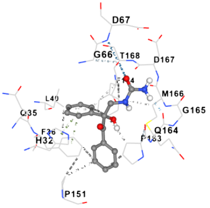
The synthesized compounds were subjected to molecular docking with the E.coli DsbA in complex with N-(2-fluorophenyl)-5-methylisoxazole-3-carboxamide structure, obtained from Protein Data Bank (PDB) (ID = 8DN0) was used in order to evaluate the synthetic chemicals' binding affinity inside the active site of the E.coli DsbA enzyme.
Compound (3) had the most advantageous docking score of -7.5 kcal/mol. Compound (4) came in second with a docking score of-7.1 kcal/mol beneficial relationships with E. Coli DsbA for more info see Table 7 blew. The produced compounds demonstrated adequate binding to the E. Coli DsbA enzyme.
Table 7. Molecular Docking result of the synthesized compounds.

Compound

Cavity size (Angstrom cubed (Å3)

Docking score (kcal/mol)

Interaction with amino acid residues

H-bonds

Hydrophobic

Native ligand

926

-6.7

HIS32,

PHE36

2

926

-6.5

HIS32,

PHE36

3

926

-7.5

HIS32, PHE36, THR161, PRO163

PHE174, LEU40,

4

926

-7.1

HIS32, THR168

PHE174

Furthermore, through a number of π-π and electrostatic interactions involving HIS32, GLN35, PHE36, LEU40, VAL150, PRO151, ALA152, LEU161 ASN162, PRO163, GLN164, GLY165, MET166, THR168, MET171, & PHE174 amino acids and hydrogen bonding between oxygen and nitrogen atoms, both compounds bonded well into the E. Coli DsbA active site. For more information see Figure 8 blew (Annex –III).
4. Conclusion
This study effectively synthesized three derivatives of 2-hydroxy-1, 2-diphenylethanone with considerable antibacterial activity. The highest activity of the three synthesized compounds against Pseudomonas aeruginosa MDR1, Shigella soneii 1, and Shigella boydii D13629 is 10µg/mL by the compound (3). Interestingly, compound (4) demonstrates greater ZOI than the standard Griseofulvin against Penicillium funiculosum NCTC 287 strains (15.0mm) and has demonstrated similar activity against Aspergillus Niger ATCC 6275 (15.0mm). Furthermore, the produced compounds showed advantageous potential interactions with the E. Coli DsbA enzyme's active site, which is essential for bacterial protein maturation and a major participant in bacterial protein biosynthesis.
Abbreviations

ADMET

Absorption, Distribution, Metabolism, Excretion and Toxicity

AMR

Anti-Microbial Resistance

DMSO

Dimethyl Sulfoxide

MIC

Minim Inhibitatory Concentration

PDB

Protein Data Bank

TLC

Thin Layer Chromatography

UV

Ultra Violet

ZOI

Zone of Inhibition

Author Contributions
Temesgen Kusse: Conceptualization, Project administration, Writing – original draft
Daniel Bisrat: Supervision, Writing – review & editing
Kaleab Asres: Validation
Avijit Mazumder: Validation
Data Availability Statement
The datasets generated during the current study are available from the corresponding author on reasonable request.
Conflicts of Interest
The authors declare no conflicts of interest.
Appendix
Appendix I: TLC
Figure 3. Normal Phase TLC of the synthesized compounds 2, 3, & 4 and starting compound 1 viewed under UV 254 using solvent system HE/CF (2: 1).
Appendix II: NMR
1H-NMR data for the synthesized compound (3)
Key: H2’, 4’ & H2’’, 4’’ protons of aromatic rings, -NH2 & NH proton of urea, H-1 methylene proton,

Download: Download full-size image

Figure 4. 1H-NMR data for the synthesized compound (3).
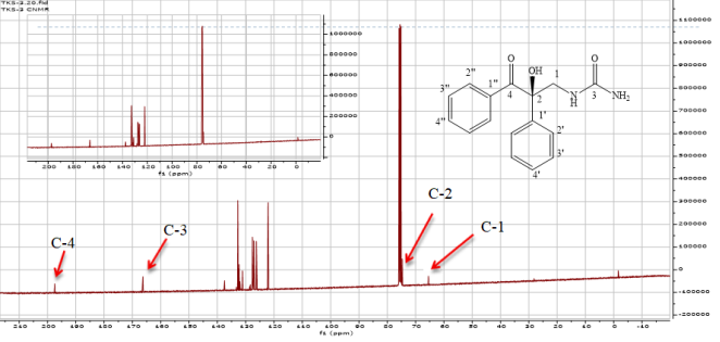
13C-NMR data for the synthesized compound 3
Figure 5. 13C-NMR data for the synthesized compound (3).
Key: C-4 Carbon of carbonyl Carbon, C-3 Carbon of carbonyl Carbon of Urea C-2 4 quaternary Carbon, C-1Methylene carbon, and peak from 120 -140 are aromatic Carbone.
1H-NMR data for the synthesized compound 4
Figure 6. 1H-NMR data for the synthesized compound (4).
Key: =CH protons of aromatic rings, -OH Hydroxyl proton, H-3&4 methylene proton Carbon connected to Nitrogen and H- 1&2 methylene proton.
13C-NMR data for the synthesized compound 4
Figure 7. 13C-NMR data for the synthesized compound (4).
Key: C-6 Carbon of carbonyl Carbon, C=C aromatic Carbone, C-5 quaternary Carbon, C-4 Methylene carbon next to chiral center, C-3 Methylene carbon next to Nitrogen, C-1&2 Methylene carbon.
Appendix III: CB-Dock Image of the Synthesized Compound 2 Complexes With E. coli DsbA
Figure 8. 3-(Diethyl amino)-2-hydroxy-1, 2-diphenylpropan-1-one complexes with E. coli DsbA.

References
[1] R. L. Berkelman, R. T. Bryan, M. T. Osterholm, J. W. LeDuc, and J. M. Hughes, “Infectious Disease Surveillance: a Crumbling Foundation,” Science (80-.)., vol. 264, no. 5157, pp. 368–370, Apr. 1994.
[2] A. Rathore, S. Sharma, and M. Sharma, “Challenges in Infectious Disease Management,” in Sustainable Nanomaterials for Treatment and Diagnosis of Infectious Diseases, John Wiley & Sons, Ltd, 2025, pp. 51–79.
[3] I. N. Okeke et al., “The scope of the antimicrobial resistance challenge,” Lancet, vol. 403, no. 10442, 2024, pp. 2426–2438.
[4] J. M. Pieniążek et al., “Overview Of The Main Bacterial Infections In Humans,” Qual. Sport, vol. 23, p. 54786, Sep. 2024.
[5] S. K. Sarsamkar and N. S. Gramopadhye, “Characterization and Antimicrobial Activity of New Cobalt-Nickel-Copper (Ii) Complexes with Benzoin and 3-Amino Benzoic Acid,” SSRN Electron. J., no. Ii, 2021.
[6] N. Yaylı et al., “Molecular docking, synthesis and biological evaluation (enzyme inhibition, antimicrobial and antioxidant) of methoxy benzoin/benzil/stilbenoid derivatives,” Org. Commun., vol. 15, no. 2, pp. 129–147, 2022.
[7] C. G. Fernández, “Benzoin, a resin produced by Styrax trees in North Sumatra Province, Indonesia,” For. Prod. livelihoods Conserv. case Stud. non-timber For. Prod. Syst., vol. 1, p. 148, 2004.
[8] H. N. Atia Sharif, R. Rehman, A. Mushtaq, and U. A. Rashid, “Review on bioactive potential of Benzoin resin,” Int. J. Chem. Biochem. Sci, vol. 10, pp. 106–110, 2016.
[9] K. Buran, “Metal complexes of sulfamethazine/benzoin-based Schiff base ligand: synthesis, characterization, DFT calculations, and antimicrobial activities,” Turkish J. Biol., vol. 49, no. SI-1, pp. 118–126, 2025.
[10] R. Babu Kothapalli, B. Anusha, M. Naveen Kumar, P. S. Singh, S. Ramakrishna, and U. Venkata Subba Reddy, “Deracemization of Benzoin and its Derivatives via Kinetic, Dynamic Kinetic, Aerobic Oxidative Kinetic, and Reagent‐mediated resolution,” Chem. Asian J., p. e202401693, 2025.
[11] K. Keenan, J. Silva Corrêa, L. Sringernyuang, S. Nayiga, and C. I. R. Chandler, “The social burden of antimicrobial resistance: what is it, how can we measure it, and why does it matter?,” JAC-Antimicrobial Resist., vol. 7, no. 2, pp. 1–10, 2025.
[12] WHO, WHO bacterial priority pathogens list, 2024.
[13] R. Nassar, G. L. Dignon, R. M. Razban, and K. A. Dill, “The Protein Folding Problem: The Role of Theory,” Journal of Molecular Biology, vol. 433, no. 20. Academic Press, p. 167126, Oct. 01, 2021.
[14] C. Santos-Martin et al., “Structural bioinformatic analysis of DsbA proteins and their pathogenicity associated substrates,” Computational and Structural Biotechnology Journal, vol. 19. Elsevier, pp. 4725–4737, Jan. 01, 2021.
[15] M. S. Iorungwa and R. A. Wuana, “Original Research article Synthesis, Characterization, Kinetics, Thermodynamic and Antimicrobial Studies of Fe (III), Cu (II), Zn (II), N, N ’ -Bis (2- hydroxy-1, 2-diphenylethanone) ethylenediamine Complexes,” no. May, 2019.
[16] F. F. Blicke, “The Mannich Reaction,” Org. React., vol. 1, pp. 303–341, 1942.
[17] J. H. Jorgensen and J. D. Turnidge, “Susceptibility test methods: dilution and disk diffusion methods,” Man. Clin. Microbiol., pp. 1253–1273, 2015.
[18] O. Daliri Shamsabadi, “Investigation of Antimicrobial Effect and Mechanical Properties of Modified Starch Films, Cellulose Nanofibers, and Citrus Essential Oils by Disk Diffusion Method,” Asian J. Green Chem., vol. 8, no. 1, pp. 1–14, 2024.
[19] R. M. Humphries et al., “CLSI methods development and standardization working group best practices for evaluation of antimicrobial susceptibility tests,” J. Clin. Microbiol., vol. 56, no. 4, pp. 10–1128, 2018.
[20] C. D. Steward et al., “Characterization of clinical isolates of Klebsiella pneumoniae from 19 laboratories using the National Committee for Clinical Laboratory Standards extended-spectrum β-lactamase detection methods,” J. Clin. Microbiol., vol. 39, no. 8, pp. 2864–2872, 2001.
[21] I. Wiegand, K. Hilpert, and R. E. W. Hancock, “Agar and broth dilution methods to determine the minimal inhibitory concentration (MIC) of antimicrobial substances,” Nat. Protoc., vol. 3, no. 2, pp. 163–175, 2008.
[22] O. Trott and A. J. Olson, “AutoDock Vina: Improving the speed and accuracy of docking with a new scoring function, efficient optimization, and multithreading,” J. Comput. Chem., vol. 31, no. 2, pp. 455–461, Jan. 2010.
[23] J. Dong et al., “ADMETlab: a platform for systematic ADMET evaluation based on a comprehensively collected ADMET database,” J. Cheminform., vol. 10, pp. 1–11, 2018.
[24] A. Garrido, A. Lepailleur, S. M. Mignani, P. Dallemagne, and C. Rochais, “hERG toxicity assessment: Useful guidelines for drug design,” Eur. J. Med. Chem., vol. 195, p. 112290, 2020.
[25] W. G.. H. B., “E.coli DsbA in complex with N-(2-fluorophenyl)-5-methylisoxazole-3-carboxamide,” Jun. 2023.
Cite This Article
  • APA Style

    Kusse, T., Bisrat, D., Asres, K., Mazumder, A. (2026). Synthesis, In vitro Antimicrobial, and In Silico Studies of 2-Hydroxy-1, 2-Diphenylethanone Derivatives. World Journal of Health Services Research, 1(1), 1-13. https://doi.org/10.11648/j.wjhsr.20260101.11

    Copy | Download

    ACS Style

    Kusse, T.; Bisrat, D.; Asres, K.; Mazumder, A. Synthesis, In vitro Antimicrobial, and In Silico Studies of 2-Hydroxy-1, 2-Diphenylethanone Derivatives. World J. Health Serv. Res. 2026, 1(1), 1-13. doi: 10.11648/j.wjhsr.20260101.11

    Copy | Download

    AMA Style

    Kusse T, Bisrat D, Asres K, Mazumder A. Synthesis, In vitro Antimicrobial, and In Silico Studies of 2-Hydroxy-1, 2-Diphenylethanone Derivatives. World J Health Serv Res. 2026;1(1):1-13. doi: 10.11648/j.wjhsr.20260101.11

    Copy | Download

  • @article{10.11648/j.wjhsr.20260101.11,
      author = {Temesgen Kusse and Daniel Bisrat and Kaleab Asres and Avijit Mazumder},
      title = {Synthesis, In vitro Antimicrobial, and In Silico Studies of 
    2-Hydroxy-1, 2-Diphenylethanone Derivatives},
      journal = {World Journal of Health Services Research},
      volume = {1},
      number = {1},
      pages = {1-13},
      doi = {10.11648/j.wjhsr.20260101.11},
      url = {https://doi.org/10.11648/j.wjhsr.20260101.11},
      eprint = {https://article.sciencepublishinggroup.com/pdf/10.11648.j.wjhsr.20260101.11},
      abstract = {Infectious diseases place a significant impact on the healthcare system. It causes 7.7 million fatalities each year, with a disproportionately high burden in sub-Saharan Africa. Drug resistant pathogens attributed to 4.95 million and 1.27 million are illnesses of bacteria that are resistant to the current medications. Therefore, there is an urgent need for new, safe, and effective compounds to combat antimicrobial resistance. The aim of this study was to synthesize new compounds derived from 2-hydroxy-1, 2-diphenylethanone using the Mannich reaction and evaluate their antimicrobial activities against 26 bacterial strains and 4 fungal strains. Three compounds were successfully synthesized and their structures were confirmed as 3 (diethylamino)-2-hydroxy-1, 2-diphenylpropan-1-one (2) 1-(2-hydroxy-3-oxo-2, 3 diphenylpropyl) urea (3), and 2-hydroxy-1, 2-diphenyl-3-(piperidin-1-yl) propan-1-one (4), using  1H and 13C-NMR spectroscopy. All the synthesized compounds exhibited broad-spectrum antibacterial activity. Compound (3) demonstrated the highest activity, with MIC of 10 µg/mL against Shigella sonnei 1, Shigella boydii D13629, and Pseudomonas aeruginosa MDR1 and compound (4) demonstrated the highest antifungal activity, with MIC of 200 µg/mL against Candida albicans ATCC 10231, Aspergillus niger ATCC 6275, Penicillium funiculosum NCTC 287, and Penicillium notatum ATCC 11625. Molecular docking showed the compounds interact favorably with conserved residues in the binding site of E. coli DsbA (PDB ID: 8DN0) through hydrogen bonding and hydrophobic interactions, with docking scores of –7.5 kcal/mol compound (3), –7.1 kcal/mol compound (4), and –6.5kcal/mol compound (2). This research suggests verify the molecular docking results experimentally and biological activities, such as antiviral, anticancer, anti-inflammatory, and antioxidant properties and evaluate the safety profile of the produced chemicals.},
     year = {2026}
    }
    

    Copy | Download

  • TY  - JOUR
    T1  - Synthesis, In vitro Antimicrobial, and In Silico Studies of 
    2-Hydroxy-1, 2-Diphenylethanone Derivatives
    AU  - Temesgen Kusse
    AU  - Daniel Bisrat
    AU  - Kaleab Asres
    AU  - Avijit Mazumder
    Y1  - 2026/09/09
    PY  - 2026
    N1  - https://doi.org/10.11648/j.wjhsr.20260101.11
    DO  - 10.11648/j.wjhsr.20260101.11
    T2  - World Journal of Health Services Research
    JF  - World Journal of Health Services Research
    JO  - World Journal of Health Services Research
    SP  - 1
    EP  - 13
    PB  - Science Publishing Group
    UR  - https://doi.org/10.11648/j.wjhsr.20260101.11
    AB  - Infectious diseases place a significant impact on the healthcare system. It causes 7.7 million fatalities each year, with a disproportionately high burden in sub-Saharan Africa. Drug resistant pathogens attributed to 4.95 million and 1.27 million are illnesses of bacteria that are resistant to the current medications. Therefore, there is an urgent need for new, safe, and effective compounds to combat antimicrobial resistance. The aim of this study was to synthesize new compounds derived from 2-hydroxy-1, 2-diphenylethanone using the Mannich reaction and evaluate their antimicrobial activities against 26 bacterial strains and 4 fungal strains. Three compounds were successfully synthesized and their structures were confirmed as 3 (diethylamino)-2-hydroxy-1, 2-diphenylpropan-1-one (2) 1-(2-hydroxy-3-oxo-2, 3 diphenylpropyl) urea (3), and 2-hydroxy-1, 2-diphenyl-3-(piperidin-1-yl) propan-1-one (4), using  1H and 13C-NMR spectroscopy. All the synthesized compounds exhibited broad-spectrum antibacterial activity. Compound (3) demonstrated the highest activity, with MIC of 10 µg/mL against Shigella sonnei 1, Shigella boydii D13629, and Pseudomonas aeruginosa MDR1 and compound (4) demonstrated the highest antifungal activity, with MIC of 200 µg/mL against Candida albicans ATCC 10231, Aspergillus niger ATCC 6275, Penicillium funiculosum NCTC 287, and Penicillium notatum ATCC 11625. Molecular docking showed the compounds interact favorably with conserved residues in the binding site of E. coli DsbA (PDB ID: 8DN0) through hydrogen bonding and hydrophobic interactions, with docking scores of –7.5 kcal/mol compound (3), –7.1 kcal/mol compound (4), and –6.5kcal/mol compound (2). This research suggests verify the molecular docking results experimentally and biological activities, such as antiviral, anticancer, anti-inflammatory, and antioxidant properties and evaluate the safety profile of the produced chemicals.
    VL  - 1
    IS  - 1
    ER  - 

    Copy | Download

Author Information
  • Abstract
  • Keywords
  • Document Sections

    1. 1. Introduction
    2. 2. Methods
    3. 3. Result and Discussion
    4. 4. Conclusion
    Show Full Outline
  • Abbreviations
  • Author Contributions
  • Data Availability Statement
  • Conflicts of Interest
  • Appendix
  • References
  • Cite This Article
  • Author Information