
Laser in situ keratomileusis (LASIK) provides predictable refractive outcomes, but clinically silent epithelial basement membrane dystrophy (EBMD) may become apparent during flap creation and lead to epithelial sloughing, delayed epithelial recovery, and postoperative ocular surface instability. This case report describes the management of a 30-year-old man with bilateral moderate myopia and astigmatism who underwent microkeratome-assisted LASIK after a routine preoperative examination showed no corneal epithelial abnormalities. The baseline Ocular Surface Disease Index score was 6, tear breakup time was 7 seconds in the right eye and 8 seconds in the left eye, and anterior-segment optical coherence tomography showed tear meniscus heights of 304 and 299 micrometers, respectively. During flap repositioning, the epithelium demonstrated abnormal mobility, folding, and focal sloughing. On postoperative day 1, the right eye had uncorrected visual acuity of 0.6-0.7, diffuse epithelial edema, and a central epithelial defect; the left eye had uncorrected visual acuity of approximately 1.0. Subconjunctival dexamethasone was administered because of the risk of diffuse lamellar keratitis. At 1 week, persistent grade 1-2 epitheliopathy and early epithelial ingrowth were observed. Treatment was supplemented with three weekly bilateral subconjunctival injections of 1% low-molecular-weight sodium hyaluronate, followed by monthly injections, together with topical corticosteroids and hyaluronic-acid lubricants. During follow-up, epithelial staining decreased, epithelial morphology on optical coherence tomography became more uniform, visual acuity remained stable, and subjective discomfort improved. At 2 months, the Ocular Surface Disease Index score decreased to 3, Schirmer test values increased to 26 and 28 mm, tear breakup time increased to 9 and 10 seconds, and tear meniscus height increased to 476 and 504 micrometers. This case suggests that multimodal regenerative therapy including subconjunctival low-molecular-weight sodium hyaluronate may support corneal epithelial recovery after intraoperative manifestation of EBMD during LASIK; controlled studies are required to determine efficacy.
| Published in | Science Journal of Clinical Medicine (Volume 15, Issue 2) |
| DOI | 10.11648/j.sjcm.20261502.14 |
| Page(s) | 21-26 |
| Creative Commons |
This is an Open Access article, distributed under the terms of the Creative Commons Attribution 4.0 International License (http://creativecommons.org/licenses/by/4.0/), which permits unrestricted use, distribution and reproduction in any medium or format, provided the original work is properly cited. |
| Copyright |
Copyright © The Author(s), 2026. Published by Science Publishing Group |
LASIK, Epithelial Basement Membrane Dystrophy, Corneal Epithelium, Hyaluronic Acid, Sodium Hyaluronate, Epithelial Sloughing, Ocular Surface, Case Report
Parameter | OD | OS |
|---|---|---|
UDVA/BCDVA | 0.1/1.0 | 0.1/1.0 |
Autokeratorefractometry, D | -5.50 -1.00 x 170° | -5.50 -0.75 x 175° |
Spherical equivalent, D | -6.00 | -6.12 |
Keratometry K1/K2, D @ axis | 43.46 @ 173° / 44.47 @ 83° | 43.31 @ 176° / 44.27 @ 86° |
Schirmer test, mm | 20 | 15 |
Central pachymetry, µm | 540 | 543 |
TBUT, s | 7 | 8 |
Meibomian gland loss, upper/lower lid,% | 5/13.7 (grade 1) | 11.4/22.9 (grade 1) |
Tear meniscus height by AS-OCT, µm | 304 | 299 |
AS-OCT | Anterior-segment Optical Coherence Tomography |
BCDVA | Best-corrected Distance Visual Acuity |
EBMD | Epithelial Basement Membrane Dystrophy |
LASIK | Laser in Situ Keratomileusis |
LMW-NaHA | Low-molecular-weight Sodium Hyaluronate |
OD | Right Eye |
OS | Left Eye |
OSDI | Ocular Surface Disease Index |
TBUT | Tear Breakup Time |
UDVA | Uncorrected Distance Visual Acuity |
| [1] | Sahay P, Bafna RK, Reddy JC, Vajpayee RB, Sharma N. Complications of laser-assisted in situ keratomileusis. Indian Journal of Ophthalmology. 2021; 69(7): 1658-1669. |
| [2] | Laibson PR. Microcystic corneal dystrophy. Trans Am Ophthalmol Soc. 1976; 74: 488–531. |
| [3] | Labbé A, Nicola RD, Dupas B, Auclin F, Baudouin C. Epithelial basement membrane dystrophy: evaluation with the HRT II Rostock Cornea Module. Ophthalmology. 2006 Aug; 113(8): 1301-8. |
| [4] | Semak GR. Activation of Regeneration and Hyaluronic Acid in Corneal Dystrophy. Minsk: Belarusian State Medical University; 2023. 171 p. |
| [5] | Dastgheib A, Thomas E, Manche E. Sloughing of corneal epithelium and wound healing complications associated with laser in situ keratomileusis in patients with epithelial basement membrane dystrophy. American Journal of Ophthalmology. 2000; 130(3): 297-303. |
| [6] | Pérez-Santonja JJ, Galal A, Cardona C, Artola A, Ruíz-Moreno JM, Alió JL. Severe corneal epithelial sloughing during laser in situ keratomileusis as a presenting sign for silent epithelial basement membrane dystrophy. Journal of Cataract & Refractive Surgery. 2005; 31(10): 1932-1937. |
| [7] | Surnina ZV, Sukhanova EV, Gamidov AA. Clinical manifestation of corneal epithelial dystrophy after LASIK. Medical Council. 2022; 16(23): 275-283. (In Russian). |
| [8] | Wu PY, Tsui MC, Chang CK, Chang HW, Chen WL. Epithelial basement membrane dystrophy after femtosecond laser-assisted LASIK successfully treated with in vivo confocal microscopy-assisted photorefractive keratectomy. Journal of Cataract & Refractive Surgery. 2020; 46(12): e17-e19. |
| [9] | Semak GR. Clinical and Experimental Substantiation of Pathogenetic Methods for Treating Chronic Dystrophic Corneal Diseases [doctoral dissertation]. Minsk; 2022. 222 p. (In Russian). |
| [10] | Semak G, Ryndova D, Smiantsina A, Kastsiukovich Y. Restoration of ocular surface homeostasis after excimer laser correction. Ophthalmology. Eastern Europe. 2023; 13(2): 97-109. |
| [11] | Zhu J, Tang X, Ho CT, Huang Q. Applications and delivery mechanisms of hyaluronic acid used for topical/transdermal delivery. International Journal of Pharmaceutics. 2020; 578: 119127. |
| [12] | Woreta FA, Davis GW, Bower KS. LASIK and surface ablation in corneal dystrophies. Survey of Ophthalmology. 2015; 60(2): 115-122. |
| [13] | Nair S, Kaur M, Sharma N, Titiyal JS. Refractive surgery and dry eye - An update. Indian Journal of Ophthalmology. 2023; 71(4): 1105-1114. |
| [14] | Kim BK, Chung YT. Clinical outcomes of epithelial basement membrane dystrophy after keratorefractive lenticule extraction. Journal of Cataract & Refractive Surgery. 2024; 50(6): 558-564. |
| [15] | Grauvogl V, Mayer WJ, Siedlecki J, Mohr N, Dirisamer M, Priglinger SG, Kassumeh S, Luft N. Corneal Epithelial Wavefront Error as a Novel Diagnostic Marker for Epithelial Basement Membrane Dystrophy. Life. 2024; 14(9): 1188. |
APA Style
Semak, G. R., Remezava, V. I., Markava, Y. V., Krishtopenko, G. V. (2026). Potential for Corneal Recovery During Nonstandard Rehabilitation After LASIK. Science Journal of Clinical Medicine, 15(2), 21-26. https://doi.org/10.11648/j.sjcm.20261502.14
ACS Style
Semak, G. R.; Remezava, V. I.; Markava, Y. V.; Krishtopenko, G. V. Potential for Corneal Recovery During Nonstandard Rehabilitation After LASIK. Sci. J. Clin. Med. 2026, 15(2), 21-26. doi: 10.11648/j.sjcm.20261502.14
AMA Style
Semak GR, Remezava VI, Markava YV, Krishtopenko GV. Potential for Corneal Recovery During Nonstandard Rehabilitation After LASIK. Sci J Clin Med. 2026;15(2):21-26. doi: 10.11648/j.sjcm.20261502.14
@article{10.11648/j.sjcm.20261502.14,
  author = {Galina Romanovna Semak and Volha Ivanovna Remezava and Yuliya Vladimirovna Markava and Gleb Valerievich Krishtopenko},
  title = {Potential for Corneal Recovery During Nonstandard Rehabilitation After LASIK},
  journal = {Science Journal of Clinical Medicine},
  volume = {15},
  number = {2},
  pages = {21-26},
  doi = {10.11648/j.sjcm.20261502.14},
  url = {https://doi.org/10.11648/j.sjcm.20261502.14},
  eprint = {https://article.sciencepublishinggroup.com/pdf/10.11648.j.sjcm.20261502.14},
  abstract = {Laser in situ keratomileusis (LASIK) provides predictable refractive outcomes, but clinically silent epithelial basement membrane dystrophy (EBMD) may become apparent during flap creation and lead to epithelial sloughing, delayed epithelial recovery, and postoperative ocular surface instability. This case report describes the management of a 30-year-old man with bilateral moderate myopia and astigmatism who underwent microkeratome-assisted LASIK after a routine preoperative examination showed no corneal epithelial abnormalities. The baseline Ocular Surface Disease Index score was 6, tear breakup time was 7 seconds in the right eye and 8 seconds in the left eye, and anterior-segment optical coherence tomography showed tear meniscus heights of 304 and 299 micrometers, respectively. During flap repositioning, the epithelium demonstrated abnormal mobility, folding, and focal sloughing. On postoperative day 1, the right eye had uncorrected visual acuity of 0.6-0.7, diffuse epithelial edema, and a central epithelial defect; the left eye had uncorrected visual acuity of approximately 1.0. Subconjunctival dexamethasone was administered because of the risk of diffuse lamellar keratitis. At 1 week, persistent grade 1-2 epitheliopathy and early epithelial ingrowth were observed. Treatment was supplemented with three weekly bilateral subconjunctival injections of 1% low-molecular-weight sodium hyaluronate, followed by monthly injections, together with topical corticosteroids and hyaluronic-acid lubricants. During follow-up, epithelial staining decreased, epithelial morphology on optical coherence tomography became more uniform, visual acuity remained stable, and subjective discomfort improved. At 2 months, the Ocular Surface Disease Index score decreased to 3, Schirmer test values increased to 26 and 28 mm, tear breakup time increased to 9 and 10 seconds, and tear meniscus height increased to 476 and 504 micrometers. This case suggests that multimodal regenerative therapy including subconjunctival low-molecular-weight sodium hyaluronate may support corneal epithelial recovery after intraoperative manifestation of EBMD during LASIK; controlled studies are required to determine efficacy.},
 year = {2026}
}
TY  - JOUR
T1  - Potential for Corneal Recovery During Nonstandard Rehabilitation After LASIK
AU  - Galina Romanovna Semak
AU  - Volha Ivanovna Remezava
AU  - Yuliya Vladimirovna Markava
AU  - Gleb Valerievich Krishtopenko
Y1  - 2026/08/20
PY  - 2026
N1  - https://doi.org/10.11648/j.sjcm.20261502.14
DO  - 10.11648/j.sjcm.20261502.14
T2  - Science Journal of Clinical Medicine
JF  - Science Journal of Clinical Medicine
JO  - Science Journal of Clinical Medicine
SP  - 21
EP  - 26
PB  - Science Publishing Group
SN  - 2327-2732
UR  - https://doi.org/10.11648/j.sjcm.20261502.14
AB  - Laser in situ keratomileusis (LASIK) provides predictable refractive outcomes, but clinically silent epithelial basement membrane dystrophy (EBMD) may become apparent during flap creation and lead to epithelial sloughing, delayed epithelial recovery, and postoperative ocular surface instability. This case report describes the management of a 30-year-old man with bilateral moderate myopia and astigmatism who underwent microkeratome-assisted LASIK after a routine preoperative examination showed no corneal epithelial abnormalities. The baseline Ocular Surface Disease Index score was 6, tear breakup time was 7 seconds in the right eye and 8 seconds in the left eye, and anterior-segment optical coherence tomography showed tear meniscus heights of 304 and 299 micrometers, respectively. During flap repositioning, the epithelium demonstrated abnormal mobility, folding, and focal sloughing. On postoperative day 1, the right eye had uncorrected visual acuity of 0.6-0.7, diffuse epithelial edema, and a central epithelial defect; the left eye had uncorrected visual acuity of approximately 1.0. Subconjunctival dexamethasone was administered because of the risk of diffuse lamellar keratitis. At 1 week, persistent grade 1-2 epitheliopathy and early epithelial ingrowth were observed. Treatment was supplemented with three weekly bilateral subconjunctival injections of 1% low-molecular-weight sodium hyaluronate, followed by monthly injections, together with topical corticosteroids and hyaluronic-acid lubricants. During follow-up, epithelial staining decreased, epithelial morphology on optical coherence tomography became more uniform, visual acuity remained stable, and subjective discomfort improved. At 2 months, the Ocular Surface Disease Index score decreased to 3, Schirmer test values increased to 26 and 28 mm, tear breakup time increased to 9 and 10 seconds, and tear meniscus height increased to 476 and 504 micrometers. This case suggests that multimodal regenerative therapy including subconjunctival low-molecular-weight sodium hyaluronate may support corneal epithelial recovery after intraoperative manifestation of EBMD during LASIK; controlled studies are required to determine efficacy.
VL  - 15
IS  - 2
ER  -
Department of Ophthalmology, Branch of Open Joint Stock Company Minsk Tractor Works - Medical Center MTZ, Minsk, Belarus
Department of Ophthalmology, Branch of Open Joint Stock Company Minsk Tractor Works - Medical Center MTZ, Minsk, Belarus
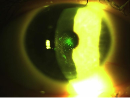
Figure 1. Fluorescein-stained anterior segment of the right eye on postoperative day 1 after LASIK, showing a central epithelial defect.
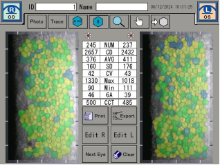
Figure 2. Specular endothelial microscopy of both eyes. No clinically significant endothelial abnormalities were identified.
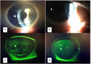
Figure 3. Anterior segment 1 month after LASIK. The right eye (A, C) shows more pronounced fluorescein staining, consistent with weaker epithelial adhesion to the basement membrane. The left eye (B, D) shows less staining and a more favorable healing pattern.
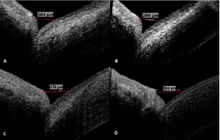
Figure 4. Tear meniscus height before surgery (A, B) and 2 months after surgery (C, D). Measurements increased from 304 and 299 µm to 476 and 504 µm in the right and left eyes, respectively.
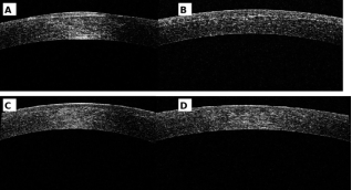
Figure 5. Anterior-segment OCT of both corneas on postoperative day 1 (A, B) and at 2 months (C, D), demonstrating improved epithelial regularity during follow-up.

Information



 Share This Article
 Twitter
 Linked In
 Facebook
 Copy
Copy Download