This case study presents a 2-month old male with Duane’s type I syndrome and bilateral severe to profound sensorineural hearing loss. The patient was referred to audiology after failing newborn hearing screen. Audiology testing revealed elevated thresholds, particularly in the left ear. Subsequent imaging demonstrated abnormal cochlear and vestibular structures, indicating an incomplete partition type I. Genetic screening for common deafness-related genes, an electrocardiogram, and infection screening for cytomegalovirus were all negative. The patient was subsequently diagnosed with left Duane’s syndrome type I at 11 months. He had a normal perinatal period and birth, with the exception of a hematoma detected around 8 weeks into gestation, which resolved without further complications. The mother received a Coronavirus-disease (COVID) vaccine during her first trimester. Family history is unremarkable for auditory or ophthalmological conditions. Duane’s retraction syndrome (DRS) is a rare form of strabismus. There is limited literature on its association with hearing loss and accompanying auditory abnormalities. We hope that this can gain further insight and provide clues to the pathogenesis and aberrant embryogenesis in this condition. Auditory screening is also important especially in settings where newborn hearing evaluation is not routine.
This is an Open Access article, distributed under the terms of the Creative Commons Attribution 4.0 International License (http://creativecommons.org/licenses/by/4.0/), which permits unrestricted use, distribution and reproduction in any medium or format, provided the original work is properly cited.
DRS is a rare congenital eye movement disorder characterized by limited horizontal eye movement, globe retraction, and narrowing of the palpebral fissure during attempted adduction.
[1]
Kekunnaya R, Negalur M. Duane retraction syndrome: causes, effects and management strategies. Clin Ophthalmol. 2017 Oct 30; 11: 1917-1930.
Kocamaz M, Aygit ED, Inal A, Ocak OB, Cicek U, Gokyigit B. Duane Retraction Syndrome and Accompanying Ocular Abnormalities. Beyoglu Eye J. 2019 Feb 20; 4(1): 28-31.
Guo Y, Zhang Q, Zhang T, Guo L, Liu S, Zhao K, Zhang W. Magnetic Resonance Imaging Findings in Patients With Duane Retraction Syndrome. J Neuroophthalmol. 2024 Mar 1; 44(1): 101-106.
DRS typically presents unilaterally, most commonly affecting the left eye, and is often diagnosed during early childhood due to abnormal eye movements or compensatory head posture
[1]
Kekunnaya R, Negalur M. Duane retraction syndrome: causes, effects and management strategies. Clin Ophthalmol. 2017 Oct 30; 11: 1917-1930.
Kocamaz M, Aygit ED, Inal A, Ocak OB, Cicek U, Gokyigit B. Duane Retraction Syndrome and Accompanying Ocular Abnormalities. Beyoglu Eye J. 2019 Feb 20; 4(1): 28-31.
Although DRS primarily affects ocular motility, it can be associated with other systemic or neurological abnormalities, including auditory defects.
[7]
Weir FW, Kreicher KL, Hatch JL, Nguyen SA, Meyer TA. Audiologic and otologic phenotype in children with Duane's Retraction Syndrome: A rare ophthalmologic disorder. Int J Pediatr Otorhinolaryngol. 2016 Oct; 89: 154-8.
[8]
Ro A, Chernoff G, MacRae D, Orton RB, Cadera W. Auditory function in Duane's retraction syndrome. Am J Ophthalmol. 1990 Jan 15; 109(1): 75-8.
Sevik O, Akdogan O, Gocmen ES, Ozcan KM, Yazar Z, Dere H. Auditory brainstem response and otoacoustic emissions in Duane retraction syndrome. Int J Pediatr Otorhinolaryngol. 2008 Aug; 72(8): 1167-70.
Hearing loss in individuals with DRS may occur as part of broader congenital syndromes or due to developmental anomalies affecting cranial nerves and related structures. The coexistence of DRS and hearing impairment has been reported in several syndromic conditions, suggesting a shared developmental origin involving neural crest cells or embryologic cranial nerve pathways.
Understanding the relationship between DRS and hearing loss is important for early diagnosis and multidisciplinary management. Early recognition of associated auditory deficits allows timely audiological evaluation and intervention, which is essential for speech, language, and cognitive development. This paper aims to explore the association between DRS and hearing loss, highlighting possible mechanisms, clinical implications, and the importance of comprehensive patient assessment.
2. Case Report
A two-month old male was referred to audiology after failing newborn hearing screen. His mother had observed that he responded inconsistently to sounds, with developmental milestones appearing normal except in listening and receptive language. Auditory brainstem response was performed and detected response thresholds >100dB at 1kHz and >85dB at 4kHz in both ears. Bone conduction was >5dB at 4kHz in the right and >45dB in the left ear. Testing results were suggestive of severe to profound bilateral hearing loss, more pronounced in the left ear. Despite the prescription of hearing aids, the patient’s response to sounds exhibited only marginal improvement.
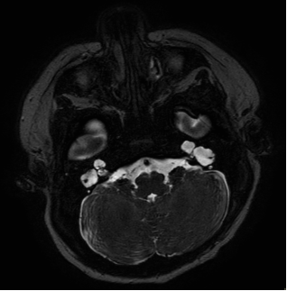
Figure 1. Cross section of magnetic imaging resonance (MRI) scan showing inner ear abnormalities as described.
A magnetic resonance imaging (MRI) scan of the internal acoustic meatus feed and wrap scan is shown in Figure 1. It revealed an enlarged and abnormal right cochlea, featuring a cystic apex and poorly defined turns superiorly. The lamina spiralis was partially preserved inferiorly. The vestibular was enlarged and continuous with a dilated, dysplastic lateral semicircular canal. The left cochlea showed enlargement and cystic characteristics with no discernible internal architecture, continuous with an abnormal, dilated vestibule and dysplastic lateral semicircular canal. The posterior and superior semi-circular canals of both ears appeared to be normal. Both internal auditory meatuses appeared slightly enlarged, and cochlear and vestibular nerves were present. There was no evidence of vestibular aqueduct enlargement of other cranial abnormalities. The overall presentation was deemed most consistent with incomplete partition type I, although atypical in the context of cochlear dilation.
Electrocardiogram results were normal, and infection screening for cytomegalovirus was negative. Genetics screening for abnormalities in genes GJB2 and GJB6, which are leading causes of autosomal recessive non-syndromic prelingual deafness, were negative. The patient had a normal perinatal period and birth, with the exception of a hematoma detected around 8 weeks into gestation, which resolved without further complications. The mother received a COVID vaccine during her first trimester. There was no family history of auditory or ophthalmological conditions.
Normal ophthalmology screens were recorded since birth, but the patient was diagnosed with left DRS type I at 11 months during a routine ophthalmology clinic. Visual acuity was 6/38 with both eyes open using Cardiff acuity cards.
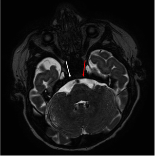
Figure 2. Cross section of MRI scan showing absent left abducens nerve (red arrow). Right abducens nerve has been labelled with a white arrow.
He had a small esotropia, with -2 restriction on abduction and narrowing of the palpebral aperture on adduction. The rest of his ocular movements were full. He had a slight left face turn. Upon dilated fundoscopy or cycloplegic refraction, the final correction was +1.5 dioptres in both eyes. Fundus examination was normal. His previous MRI was retrospectively reviewed which showed an absent left abducens nerve (Figure 2). Oculomotor nerves were normal bilaterally and lateral rectus mucles were symmetrical in both eyes.
3. Discussion
Hearing problems in DRS has been reported, with several syndromes presenting with congenital hearing loss and DRS such as Wildervanck’s cervico-oculo-acusticus syndrome, Okihiro syndrome and Bosley-Salih-Alorainy syndrome. A retrospective study of 79 patients with DRS found that 22 had some form of hearing loss.
[7]
Weir FW, Kreicher KL, Hatch JL, Nguyen SA, Meyer TA. Audiologic and otologic phenotype in children with Duane's Retraction Syndrome: A rare ophthalmologic disorder. Int J Pediatr Otorhinolaryngol. 2016 Oct; 89: 154-8.
[7]
It has been hypothesized that hearing problems are associated with DRS due to associated structural auditory malformations in the ears which occur alongside ocular malformations. Development of cranial nerves and major ear structures occur in week 4-10 of gestation, hence an insult during this period of gestation might result in abnormalities of both. There is currently limited literature on DRS with hearing loss, with most studies focusing mainly on characterising the prevalence and types of hearing loss in DRS patients. To the best of our knowledge, there has only been one other case study reporting on the cochleovestibular anatomy of ear malformations seen in Duane’s
[10]
Tachibana M, Hoshino A, Oshima W, Nishimura H, Mizukoshi O. Duane's Syndrome Associated With Crocodile Tear and Ear Malformation: A Case of Stapes Anomaly and Its Reconstruction. Arch Otolaryngol. 1984; 110(11): 761–762.
[10]
.
By describing a comprehensive audiological review and characterising the ear anatomy of a DRS patient with severe hearing loss, we hope that this can provide clues to the pathogenesis and aberrant embryogenesis of this condition. Incomplete partition type I phenotype has been reported to be due to the disruption of microvascular structures supporting the cochlear nerve.
[11]
Brotto D, Sorrentino F, Cenedese R, Avato I, Bovo R, Trevisi P, Manara R. Genetics of Inner Ear Malformations: A Review. Audiol Res. 2021 Oct 12; 11(4): 524-536.
[11]
We are unable to ascertain the significance of the uterine haematoma with regards to this. To our knowledge, several genes have been associated in DRS with hearing loss such as HOXA1 and MAFB. MAFB has been associated with inner ear abnormalities in animal studies
[12]
Chen X, Huang Y, Gao P, Lv Y, Jia D, Sun K, Han Y, Hu H, Tang Z, Ren X, Liu M. Knockout of mafba Causes Inner-Ear Developmental Defects in Zebrafish via the Impairment of Proliferation and Differentiation of Ionocyte Progenitor Cells. Biomedicines. 2021 Nov 16; 9(11): 1699.
[13]
Park JG, Tischfield MA, Nugent AA, Cheng L, Di Gioia SA, Chan WM, Maconachie G, Bosley TM, Summers CG, Hunter DG, Robson CD, Gottlob I, Engle EC. Loss of MAFB Function in Humans and Mice Causes Duane Syndrome, Aberrant Extraocular Muscle Innervation, and Inner-Ear Defects. Am J Hum Genet. 2016 Jun 2; 98(6): 1220-1227.
[14]
Choo D, Ward J, Reece A, Dou H, Lin Z, Greinwald J. Molecular mechanisms underlying inner ear patterning defects in kreisler mutants. Developmental Biology 2006 Jan; 289 (308-317).
[15]
Atay G, Tellioğlu B, Tellioğlu HT, Avcı NB, Çınar BÇ, Şekeroğlu HT. Evaluation of auditory pathways and comorbid inner ear malformations in pediatric patients with Duane retraction syndrome. Int J Pediatr Otorhinolaryngol. 2025 Jan; 188: 112207.
and there may be a role for genetic analysis although there are no other systemic abnormalities of note in our patient.
4. Conclusion
Hearing loss is associated with DRS. Our case report details the specific auditory abnormality associated. It is therefore essential to screen all DRS patients with an audiological evaluation, especially in healthcare systems where newborn hearing screen is not routine. Early diagnosis of concurrent hearing loss is crucial for timely intervention and proper audiological management to ensure proper child development. Likewise, patients with audiological abnormalities should also be considered for routine Ophthalmology examination. We are uncertain on the significance of the uterine haematoma or COVID vaccination as contributory factors, but the timing coincides with embryogenesis of the ear and eye. Further work to investigate whether there is a common progenitor origin involved in the embryogenesis of ear and eye development can help further provide insight into the pathogenesis of DRS.
Abbreviations
COVID
Coronavirus-disease
DRS
Duane’s Retraction Synd Rome
MRI
Magnetic Resonance Imaging
Author Contributions
Yan Tong Koh: Conceptualization, Formal Analysis, Project administration, Writing – original draft, Writing – review & editing
Ka Lam Janice Wu: Data curation, Formal Analysis, Writing – original draft, Writing – review & editing
Tin Chan: Investigation, Methodology, Supervision, Conceptualization
Samantha Choi: Data curation, Formal Analysis, Visualization
Conflicts of Interest
The authors declare no conflicts of interest.
References
[1]
Kekunnaya R, Negalur M. Duane retraction syndrome: causes, effects and management strategies. Clin Ophthalmol. 2017 Oct 30; 11: 1917-1930.
Kocamaz M, Aygit ED, Inal A, Ocak OB, Cicek U, Gokyigit B. Duane Retraction Syndrome and Accompanying Ocular Abnormalities. Beyoglu Eye J. 2019 Feb 20; 4(1): 28-31.
Guo Y, Zhang Q, Zhang T, Guo L, Liu S, Zhao K, Zhang W. Magnetic Resonance Imaging Findings in Patients With Duane Retraction Syndrome. J Neuroophthalmol. 2024 Mar 1; 44(1): 101-106.
Weir FW, Kreicher KL, Hatch JL, Nguyen SA, Meyer TA. Audiologic and otologic phenotype in children with Duane's Retraction Syndrome: A rare ophthalmologic disorder. Int J Pediatr Otorhinolaryngol. 2016 Oct; 89: 154-8.
[8]
Ro A, Chernoff G, MacRae D, Orton RB, Cadera W. Auditory function in Duane's retraction syndrome. Am J Ophthalmol. 1990 Jan 15; 109(1): 75-8.
Sevik O, Akdogan O, Gocmen ES, Ozcan KM, Yazar Z, Dere H. Auditory brainstem response and otoacoustic emissions in Duane retraction syndrome. Int J Pediatr Otorhinolaryngol. 2008 Aug; 72(8): 1167-70.
Tachibana M, Hoshino A, Oshima W, Nishimura H, Mizukoshi O. Duane's Syndrome Associated With Crocodile Tear and Ear Malformation: A Case of Stapes Anomaly and Its Reconstruction. Arch Otolaryngol. 1984; 110(11): 761–762.
[11]
Brotto D, Sorrentino F, Cenedese R, Avato I, Bovo R, Trevisi P, Manara R. Genetics of Inner Ear Malformations: A Review. Audiol Res. 2021 Oct 12; 11(4): 524-536.
[12]
Chen X, Huang Y, Gao P, Lv Y, Jia D, Sun K, Han Y, Hu H, Tang Z, Ren X, Liu M. Knockout of mafba Causes Inner-Ear Developmental Defects in Zebrafish via the Impairment of Proliferation and Differentiation of Ionocyte Progenitor Cells. Biomedicines. 2021 Nov 16; 9(11): 1699.
[13]
Park JG, Tischfield MA, Nugent AA, Cheng L, Di Gioia SA, Chan WM, Maconachie G, Bosley TM, Summers CG, Hunter DG, Robson CD, Gottlob I, Engle EC. Loss of MAFB Function in Humans and Mice Causes Duane Syndrome, Aberrant Extraocular Muscle Innervation, and Inner-Ear Defects. Am J Hum Genet. 2016 Jun 2; 98(6): 1220-1227.
[14]
Choo D, Ward J, Reece A, Dou H, Lin Z, Greinwald J. Molecular mechanisms underlying inner ear patterning defects in kreisler mutants. Developmental Biology 2006 Jan; 289 (308-317).
[15]
Atay G, Tellioğlu B, Tellioğlu HT, Avcı NB, Çınar BÇ, Şekeroğlu HT. Evaluation of auditory pathways and comorbid inner ear malformations in pediatric patients with Duane retraction syndrome. Int J Pediatr Otorhinolaryngol. 2025 Jan; 188: 112207.
Koh, Y. T., Wu, K. L. J., Chan, T., Choi, S. (2026). It Is Not Just the Eyes - a Description of Abnormal Vestibulocochlear Anatomy in Duane’s Retraction Syndrome (DRS). International Journal of Ophthalmology & Visual Science, 11(3), 31-34. https://doi.org/10.11648/j.ijovs.20261103.11
Koh, Y. T.; Wu, K. L. J.; Chan, T.; Choi, S. It Is Not Just the Eyes - a Description of Abnormal Vestibulocochlear Anatomy in Duane’s Retraction Syndrome (DRS). Int. J. Ophthalmol. Vis. Sci.2026, 11(3), 31-34. doi: 10.11648/j.ijovs.20261103.11
Koh YT, Wu KLJ, Chan T, Choi S. It Is Not Just the Eyes - a Description of Abnormal Vestibulocochlear Anatomy in Duane’s Retraction Syndrome (DRS). Int J Ophthalmol Vis Sci. 2026;11(3):31-34. doi: 10.11648/j.ijovs.20261103.11
@article{10.11648/j.ijovs.20261103.11,
  author = {Yan Tong Koh and Ka Lam Janice Wu and Tin Chan and Samantha Choi},
  title = {It Is Not Just the Eyes - a Description of Abnormal Vestibulocochlear Anatomy in Duane’s Retraction Syndrome (DRS)},
  journal = {International Journal of Ophthalmology & Visual Science},
  volume = {11},
  number = {3},
  pages = {31-34},
  doi = {10.11648/j.ijovs.20261103.11},
  url = {https://doi.org/10.11648/j.ijovs.20261103.11},
  eprint = {https://article.sciencepublishinggroup.com/pdf/10.11648.j.ijovs.20261103.11},
  abstract = {This case study presents a 2-month old male with Duane’s type I syndrome and bilateral severe to profound sensorineural hearing loss. The patient was referred to audiology after failing newborn hearing screen. Audiology testing revealed elevated thresholds, particularly in the left ear. Subsequent imaging demonstrated abnormal cochlear and vestibular structures, indicating an incomplete partition type I. Genetic screening for common deafness-related genes, an electrocardiogram, and infection screening for cytomegalovirus were all negative. The patient was subsequently diagnosed with left Duane’s syndrome type I at 11 months. He had a normal perinatal period and birth, with the exception of a hematoma detected around 8 weeks into gestation, which resolved without further complications. The mother received a Coronavirus-disease (COVID) vaccine during her first trimester. Family history is unremarkable for auditory or ophthalmological conditions. Duane’s retraction syndrome (DRS) is a rare form of strabismus. There is limited literature on its association with hearing loss and accompanying auditory abnormalities. We hope that this can gain further insight and provide clues to the pathogenesis and aberrant embryogenesis in this condition. Auditory screening is also important especially in settings where newborn hearing evaluation is not routine.},
 year = {2026}
}
TY  - JOUR
T1  - It Is Not Just the Eyes - a Description of Abnormal Vestibulocochlear Anatomy in Duane’s Retraction Syndrome (DRS)
AU  - Yan Tong Koh
AU  - Ka Lam Janice Wu
AU  - Tin Chan
AU  - Samantha Choi
Y1  - 2026/07/22
PY  - 2026
N1  - https://doi.org/10.11648/j.ijovs.20261103.11
DO  - 10.11648/j.ijovs.20261103.11
T2  - International Journal of Ophthalmology & Visual Science
JF  - International Journal of Ophthalmology & Visual Science
JO  - International Journal of Ophthalmology & Visual Science
SP  - 31
EP  - 34
PB  - Science Publishing Group
SN  - 2637-3858
UR  - https://doi.org/10.11648/j.ijovs.20261103.11
AB  - This case study presents a 2-month old male with Duane’s type I syndrome and bilateral severe to profound sensorineural hearing loss. The patient was referred to audiology after failing newborn hearing screen. Audiology testing revealed elevated thresholds, particularly in the left ear. Subsequent imaging demonstrated abnormal cochlear and vestibular structures, indicating an incomplete partition type I. Genetic screening for common deafness-related genes, an electrocardiogram, and infection screening for cytomegalovirus were all negative. The patient was subsequently diagnosed with left Duane’s syndrome type I at 11 months. He had a normal perinatal period and birth, with the exception of a hematoma detected around 8 weeks into gestation, which resolved without further complications. The mother received a Coronavirus-disease (COVID) vaccine during her first trimester. Family history is unremarkable for auditory or ophthalmological conditions. Duane’s retraction syndrome (DRS) is a rare form of strabismus. There is limited literature on its association with hearing loss and accompanying auditory abnormalities. We hope that this can gain further insight and provide clues to the pathogenesis and aberrant embryogenesis in this condition. Auditory screening is also important especially in settings where newborn hearing evaluation is not routine.
VL  - 11
IS  - 3
ER  -
Department of Ophthalmology, Tan Tock Seng Hospital, Singapore; Ophthalmology Department, Royal Hospital for Children & Young People, Edinburgh, United Kingdom; Princess Alexandra Eye Pavilion, Edinburgh, United Kingdom
Ophthalmology Department, Royal Hospital for Children & Young People, Edinburgh, United Kingdom; Princess Alexandra Eye Pavilion, Edinburgh, United Kingdom
Koh, Y. T., Wu, K. L. J., Chan, T., Choi, S. (2026). It Is Not Just the Eyes - a Description of Abnormal Vestibulocochlear Anatomy in Duane’s Retraction Syndrome (DRS). International Journal of Ophthalmology & Visual Science, 11(3), 31-34. https://doi.org/10.11648/j.ijovs.20261103.11
Koh, Y. T.; Wu, K. L. J.; Chan, T.; Choi, S. It Is Not Just the Eyes - a Description of Abnormal Vestibulocochlear Anatomy in Duane’s Retraction Syndrome (DRS). Int. J. Ophthalmol. Vis. Sci.2026, 11(3), 31-34. doi: 10.11648/j.ijovs.20261103.11
Koh YT, Wu KLJ, Chan T, Choi S. It Is Not Just the Eyes - a Description of Abnormal Vestibulocochlear Anatomy in Duane’s Retraction Syndrome (DRS). Int J Ophthalmol Vis Sci. 2026;11(3):31-34. doi: 10.11648/j.ijovs.20261103.11
@article{10.11648/j.ijovs.20261103.11,
  author = {Yan Tong Koh and Ka Lam Janice Wu and Tin Chan and Samantha Choi},
  title = {It Is Not Just the Eyes - a Description of Abnormal Vestibulocochlear Anatomy in Duane’s Retraction Syndrome (DRS)},
  journal = {International Journal of Ophthalmology & Visual Science},
  volume = {11},
  number = {3},
  pages = {31-34},
  doi = {10.11648/j.ijovs.20261103.11},
  url = {https://doi.org/10.11648/j.ijovs.20261103.11},
  eprint = {https://article.sciencepublishinggroup.com/pdf/10.11648.j.ijovs.20261103.11},
  abstract = {This case study presents a 2-month old male with Duane’s type I syndrome and bilateral severe to profound sensorineural hearing loss. The patient was referred to audiology after failing newborn hearing screen. Audiology testing revealed elevated thresholds, particularly in the left ear. Subsequent imaging demonstrated abnormal cochlear and vestibular structures, indicating an incomplete partition type I. Genetic screening for common deafness-related genes, an electrocardiogram, and infection screening for cytomegalovirus were all negative. The patient was subsequently diagnosed with left Duane’s syndrome type I at 11 months. He had a normal perinatal period and birth, with the exception of a hematoma detected around 8 weeks into gestation, which resolved without further complications. The mother received a Coronavirus-disease (COVID) vaccine during her first trimester. Family history is unremarkable for auditory or ophthalmological conditions. Duane’s retraction syndrome (DRS) is a rare form of strabismus. There is limited literature on its association with hearing loss and accompanying auditory abnormalities. We hope that this can gain further insight and provide clues to the pathogenesis and aberrant embryogenesis in this condition. Auditory screening is also important especially in settings where newborn hearing evaluation is not routine.},
 year = {2026}
}
TY  - JOUR
T1  - It Is Not Just the Eyes - a Description of Abnormal Vestibulocochlear Anatomy in Duane’s Retraction Syndrome (DRS)
AU  - Yan Tong Koh
AU  - Ka Lam Janice Wu
AU  - Tin Chan
AU  - Samantha Choi
Y1  - 2026/07/22
PY  - 2026
N1  - https://doi.org/10.11648/j.ijovs.20261103.11
DO  - 10.11648/j.ijovs.20261103.11
T2  - International Journal of Ophthalmology & Visual Science
JF  - International Journal of Ophthalmology & Visual Science
JO  - International Journal of Ophthalmology & Visual Science
SP  - 31
EP  - 34
PB  - Science Publishing Group
SN  - 2637-3858
UR  - https://doi.org/10.11648/j.ijovs.20261103.11
AB  - This case study presents a 2-month old male with Duane’s type I syndrome and bilateral severe to profound sensorineural hearing loss. The patient was referred to audiology after failing newborn hearing screen. Audiology testing revealed elevated thresholds, particularly in the left ear. Subsequent imaging demonstrated abnormal cochlear and vestibular structures, indicating an incomplete partition type I. Genetic screening for common deafness-related genes, an electrocardiogram, and infection screening for cytomegalovirus were all negative. The patient was subsequently diagnosed with left Duane’s syndrome type I at 11 months. He had a normal perinatal period and birth, with the exception of a hematoma detected around 8 weeks into gestation, which resolved without further complications. The mother received a Coronavirus-disease (COVID) vaccine during her first trimester. Family history is unremarkable for auditory or ophthalmological conditions. Duane’s retraction syndrome (DRS) is a rare form of strabismus. There is limited literature on its association with hearing loss and accompanying auditory abnormalities. We hope that this can gain further insight and provide clues to the pathogenesis and aberrant embryogenesis in this condition. Auditory screening is also important especially in settings where newborn hearing evaluation is not routine.
VL  - 11
IS  - 3
ER  -





 Share This Article
 Twitter
 Linked In
 Facebook
 Copy
Copy Download