Department of Stomatology and maxillofacial surgery, Ledea Bernard Ouedraogo University and Regional University Hospital Center of Ouahigouya, Ouahigouya, Burkina Faso
Department of Otolaryngology and Head and Neck Surgery, Ledea Bernard Ouedraogo University and Regional University Hospital Center of Ouahigouya, Ouahigouya, Burkina Faso
Introduction: Imaging examinations play an essential role in the diagnosis and management of stomatological and maxillofacial conditions. This study aimed to describe imaging prescription patterns in stomatology and maxillofacial surgery at the Ouahigouya Regional University Hospital Center (CHUR-OHG) from January 1 to December 31, 2024, and to assess their clinical relevance in the local context. Materials and Methods: We conducted a descriptive, retrospective, observational cross-sectional study including all patients managed in the stomatology and maxillofacial surgery department during the study period. Results: Of the 637 consultations recorded, 323 patients (50.7%) received an imaging prescription. The study population was predominantly male (67.2%; sex ratio, 2.04), with a mean age of 32 years, and most patients were from urban areas (71.8%). Infectious (44.6%) and traumatic (41.5%) conditions were the leading indications for imaging. Standard radiography (51.2%) and computed tomography (44.8%) were the most frequently prescribed modalities, together accounting for 96.0% of all prescriptions, whereas ultrasound and magnetic resonance imaging were rarely prescribed. Among standard radiographic examinations, panoramic radiography accounted for 99.4%. Overall, 87.6% of prescribed examinations were confirmed as having been performed. Conclusion: Imaging plays a central role in stomatology and maxillofacial surgery at CHUR-OHG, particularly in the management of infectious and traumatic conditions. The predominance of panoramic radiography and computed tomography reflects the main diagnostic needs of the population served. These findings provide useful baseline data to support more appropriate imaging prescriptions and to guide efforts to optimize access to and use of diagnostic imaging in the Burkinabe context.
This is an Open Access article, distributed under the terms of the Creative Commons Attribution 4.0 International License (http://creativecommons.org/licenses/by/4.0/), which permits unrestricted use, distribution and reproduction in any medium or format, provided the original work is properly cited.
Maxillofacial surgery, which emerged from stomatology, developed considerably throughout the 20th century. It now encompasses a broad range of conditions, including trauma, infections, tumors, malformations, and facial reconstruction
[1]
French Hospital Federation. Stomatology and maxillofacial surgery - hopital.fr - French Hospital Federation [Internet]. Hopital.fr. 2025.
[1]
. Medical imaging has become an indispensable component of this field, providing essential support for diagnosis, disease assessment, and treatment planning. Imaging prescriptions should therefore be based on appropriate clinical criteria to ensure a favorable benefit–risk balance, particularly in view of the potential risks associated with ionizing radiation exposure
[2]
French Society of Stomatology, Maxillofacial Surgery, and Oral Surgery (SFSCMFCO). Chossegros C, ed. Imaging Techniques in Orthognathic Surgery; Best Practice Recommendations; Paris, France; July 2017.
[2]
.
The choice of imaging modality varies according to the type of lesion and the anatomical structures involved. Standard radiography, particularly panoramic radiography (orthopantomography), is commonly used as a first-line imaging modality. Computed tomography (CT) and cone-beam computed tomography (CBCT) provide detailed assessment of osseous structures and anatomical cavities, whereas magnetic resonance imaging (MRI) is particularly suited to the evaluation of soft tissues
[3]
Bourjat P. Useful Imaging in Maxillofacial Surgery. Part Two: Practical Applications. Rev Stomatol Chir Maxillofac. 2007; 108(1): 31-45.
[3]
.
Several international studies have reported a predominance of panoramic radiography, including studies conducted in Morocco
[4]
Haouane MA. Cervicofacial Cellulitis. Experience of the Department of Maxillofacial Surgery and Stomatology, Avicenne Military Hospital, Marrakesh [Medical Dissertation]. Marrakesh: Cadi Ayyad University; 2018.
[4]
. In Burkina Faso, a survey conducted in Ouagadougou in 2012 reported the use of Blondeau and orthopantomographic views
[5]
Ganebang P. Stomatological pathology and maxillofacial surgery in private practice in Ouagadougou: a study of 385 cases [Medical Dissertation]. Ouagadougou: University of Ouagadougou; 2012.
[5]
. These findings highlight the importance of adapting imaging prescriptions to local clinical and healthcare contexts while taking into account technical and financial constraints.
To date, no systematic study has specifically examined imaging prescription patterns in stomatology and maxillofacial surgery at Ouahigouya Regional University Hospital Center (CHUR-OHG). This study therefore sought to address this gap by describing current prescription practices, analyzing the clinical indications for imaging, and comparing the observed patterns with those reported in the literature. More specifically, it aimed to provide reliable data on the concordance between prescribed imaging examinations and the conditions being investigated, with the goal of contributing to improved prescription practices and patient care in the Burkinabe context. We therefore assessed imaging prescriptions in stomatology and maxillofacial surgery at CHUR-OHG from January 1 to December 31, 2024.
2. Materials and Methods
2.1. Study Design and Period
This was a retrospective, descriptive, cross-sectional study conducted from January 1 to December 31, 2024, in the Department of Stomatology and Maxillofacial Surgery at CHUR-OHG.
2.2. Study Population
The study included all patients seen in the Department of Stomatology and Maxillofacial Surgery at CHUR-OHG between January 1 and December 31, 2024.
2.3. Inclusion and Exclusion Criteria
We included all patients who received an imaging prescription during the study period. Patients with incomplete or unusable medical records were excluded, particularly those with missing documentation of imaging prescriptions (examination requests, reports, or results) or insufficient clinical data.
2.4. Data Collection and Sources
Data were collected from consultation registers and imaging requisition forms. The variables assessed included age, sex, place of residence, clinical indication, type of imaging examination prescribed, and whether the examination was performed. Data were analyzed using SPSS, and results were summarized as frequencies and proportions.
2.5. Ethical Considerations
Data were collected while ensuring patient confidentiality and anonymity. Authorization to conduct the study was obtained from the Director General of CHUR-OHG prior to data collection.
3. Results
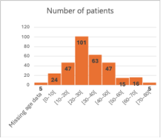
Of the 637 consultations recorded, 323 patients (50.7%) received an imaging prescription. The mean age was 32 years, with a male predominance (67.2%; sex ratio, 2.04) as Figure 1. Most patients were from urban areas (71.8%).
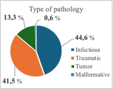
Figure 2. Distribution of imaging indications according to pathology type.
The main indications for imaging were infectious (44.6%) and traumatic (41.5%) conditions as Figure 2.
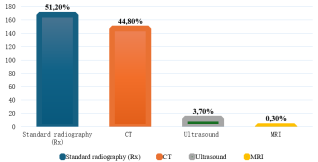
Standard radiography and computed tomography (CT) accounted for 51.2% and 44.8% of all imaging prescriptions, respectively, together representing 96.0% of prescriptions. Ultrasound and magnetic resonance imaging (MRI) were rarely prescribed as Figure 3.
Figure 3. Distribution of requested imaging examinations by type.
The imaging modality varied according to the clinical indication. For infectious conditions, standard radiography was the predominant modality, accounting for 93.1% of prescriptions. Among patients with traumatic conditions, CT was prescribed in 110 cases (82.1%) as Table 1.
Table 1. Distribution of imaging prescriptions for traumatic conditions.
Imaging exam type for traumatic conditions
Number
Percentage (%)
Standard radiography (panoramic)
23
17.2
CT
110
82.1
MRI
1
0.7
Ultrasound
0
0.0
For tumoral conditions, CT was prescribed in 31 patients (64.6%) as Table 2. Standard radiography was prescribed in all patients with malformative conditions (100%). All standard radiographic examinations were extraoral. Panoramic radiography accounted for nearly all extraoral radiographs (99.4%), while the Blondeau view accounted for 0.6%.
Table 2. Distribution of imaging prescriptions for tumoral conditions.
Imaging exam type for tumoral conditions
Number
Percentage (%)
Standard radiography (panoramic)
9
18.8
CT
31
64.6
Ultrasound
8
16.7
MRI
0
0.0
The craniofacial region was the most frequently targeted anatomical region, accounting for 312 examinations (96.6% of all imaging prescriptions), whereas the cervicofacial region was targeted in 4 cases (1.2%) as Table 3.
Overall, 87.6% of prescribed imaging examinations were confirmed as having been performed.
Table 3. Distribution of imaging examinations by anatomical region.
Region explored
Frequency
Percentage (%)
Craniofacial
312
96.6
Cervicofacial
4
1.2
Cranioencephalic
1
0.3
Craniofacial + cranioencephalic
3
0.9
Craniofacial + cervicothoracic
2
0.6
Craniofacial + cranioencephalic + cervicofacial
1
0.3
4. Discussion
This study provides an overview of imaging prescription patterns in stomatology and maxillofacial surgery at CHUR-OHG. Among the 637 consultations reviewed, 50.7% resulted in an imaging prescription, highlighting the important role of imaging in the diagnosis and management of maxillofacial conditions. This relatively high proportion may partly reflect the referral nature of the institution, which is likely to receive patients with more complex conditions.
The mean age was 32 years, consistent with the findings of Ouedraogo (32.6 years)
[6]
Ouedraogo W. Cervicofacial Cellulitis in the Department of Odontostomatology at CHUR-OHG in Burkina Faso: A Report on 40 Cases [Medical Dissertation]. Ouahigouya: Ledea Bernard Ouedraogo University; 2020.
[6]
and Haouane (33.7 years)
[4]
Haouane MA. Cervicofacial Cellulitis. Experience of the Department of Maxillofacial Surgery and Stomatology, Avicenne Military Hospital, Marrakesh [Medical Dissertation]. Marrakesh: Cadi Ayyad University; 2018.
[4]
. The predominance of young adults may be related to the epidemiological profile of conditions commonly encountered in maxillofacial practice, particularly trauma and infections. Men accounted for 67.2% of the study population, with a sex ratio of 2.04, consistent with the ratios reported by Ganebang in Ouagadougou and Lebeau in France (2.7)
[5]
Ganebang P. Stomatological pathology and maxillofacial surgery in private practice in Ouagadougou: a study of 385 cases [Medical Dissertation]. Ouagadougou: University of Ouagadougou; 2012.
[7]
Traore PD. Management of Long-Standing Maxillofacial Tumors at the National Center for Odonto-Stomatology (Pr HT) in Bamako [Medical Dissertation]. Bamako: University of Sciences, Techniques and Technologies of Bamako; 2023.
[8]
Lebeau J, Kanku V, Duroure F, Morand B, Sadek H, Raphaël B. Facial Trauma at Grenoble University Hospital: An Epidemiological Study of 961 Cases Over a 365-Day Period. Rev Stomatol Chir Maxillofac. 2006; 107(1): 23-29.
. This male predominance may partly reflect greater exposure to road traffic injuries, occupational hazards, and other traumatic risk factors, although differences in healthcare-seeking behavior may also contribute.
Infectious conditions were the leading indication for imaging (44.6%), followed by trauma (41.5%), tumors (13.3%), and malformations (0.6%). These findings are close to those reported by Savadogo (49.3% infectious and 43.0% traumatic)
[9]
Savadogo L. Epidemiology of Oral and Maxillofacial Diseases at the Ouahigouya Regional University Hospital [Medical Dissertation]. Ouahigouya: Ledea Bernard Ouedraogo University; 2025.
[9]
, but differ from Ganebang, who reported a predominance of traumatic conditions (61.8%)
[5]
Ganebang P. Stomatological pathology and maxillofacial surgery in private practice in Ouagadougou: a study of 385 cases [Medical Dissertation]. Ouagadougou: University of Ouagadougou; 2012.
[5]
. Such differences may reflect variations in patient populations, referral patterns, institutional profiles, and local epidemiological characteristics.
Standard radiography and CT accounted for 51.2% and 44.8% of prescriptions, respectively, representing 96.0% of all imaging examinations. Standard radiography was predominantly used for infectious conditions (93.1%) and consisted almost exclusively of panoramic radiography (99.4%). This finding is similar to that reported by Camara in Bamako (99.2%)
[10]
Camara F. Contribution of dental radiography to the Oral Surgery Department of CHU-CNOS: a study of 483 cases. [Medical Dissertation]. Bamako: University of Sciences, Techniques and Technologies of Bamako; 2021.
[10]
and may be explained by the broad anatomical coverage, accessibility, and relatively low cost of panoramic radiography.
CT was particularly predominant in traumatic conditions, being prescribed in 82.1% of cases. This finding is consistent with the established role of CT as the primary imaging modality for the assessment and characterization of maxillofacial trauma
[11]
Fabrega M. Imaging of Maxillofacial Trauma. Oral Maxillofac Surg Clin North Am. 2023 Aug; 35(3): 297-309.
. However, the high proportion observed in our study should also be considered in light of radiation exposure and the need for dose optimization, particularly in settings where CT availability may influence prescribing patterns
[12]
Widmann G, Schönthaler H, Tartarotti A, Degenhart G, Hörmann R, Feuchtner G, Jacobs R, Pauwels R. As low as diagnostically acceptable dose imaging in maxillofacial trauma: a reference quality approach. Dentomaxillofac Radiol. 2023 Feb; 52(3): 20220387.
Ouedraogo Z. Orbitozygomatic fractures: epidemiological, diagnostic, therapeutic and outcome aspects at CHUR-OHG. [Medical Dissertation]. Ledea Bernard Ouedraogo University; 2025.
[12, 13]
. The difference from the proportion reported by Djogbe in Lomé (33.8%) and by Traoré in Bamako may reflect variations in referral patterns, case severity, institutional practices, and access to CT
Djemi EM, N'guessan EZ, Pornan IB, Ba MAA, Yassi GD, Alexandre DOO, et al. Traumatic emergencies in stomatology and maxillofacial surgery: injury patterns, therapeutic management and outcomes. Health Sci Dis. 2023; 24(2).
[14, 15]
.
For tumoral conditions, CT was the most frequently prescribed modality (64.6%), followed by ultrasound (16.7%). This pattern reflects the complementary role of cross-sectional imaging and ultrasound in assessing lesion extent and anatomical involvement. The predominance of CT is also consistent with findings reported by Traoré. The limited use of MRI may, however, reflect its availability and accessibility in the study setting.
Malformative conditions were rare (0.6%) and were evaluated exclusively using standard radiography. The limited availability of advanced modalities such as CBCT may partly explain this pattern. Although advanced imaging can provide more detailed anatomical characterization in selected craniofacial conditions, its use remains dependent on availability, cost, and local clinical practice.
Overall, 87.6% of prescribed examinations were confirmed as having been performed, suggesting good traceability within the study setting. However, some examinations may have been performed outside the institution and were therefore not documented, potentially leading to an underestimation of the actual completion rate.
This study has several limitations. Its retrospective and descriptive design limited the completeness of available information and prevented assessment of the appropriateness of individual prescriptions or causal relationships. The single-center setting also limits generalizability. In addition, the study did not assess diagnostic yield, radiation exposure, costs, or the impact of imaging findings on clinical management.
Despite these limitations, our findings provide useful baseline data on imaging practices in stomatology and maxillofacial surgery at CHUR-OHG. The predominance of infectious and traumatic indications and the substantial use of panoramic radiography and CT highlight the major diagnostic needs of this population. Further prospective and multicenter studies should assess the appropriateness, diagnostic yield, cost-effectiveness, and clinical impact of imaging prescriptions. Such evidence could support the development of context-specific imaging guidelines and promote more rational and efficient use of diagnostic imaging in maxillofacial care.
5. Conclusions
Imaging prescriptions in stomatology and maxillofacial surgery at CHUR-OHG were mainly driven by infectious and traumatic conditions, with panoramic radiography and CT accounting for the vast majority of examinations. These findings highlight their central role in the diagnostic management of maxillofacial conditions in this setting. This study provides useful baseline data to support more appropriate imaging prescriptions and contribute to the optimization of diagnostic care in the Burkinabe context.
Abbreviations
CHUR
Regional University Hospital Center
CHUR-OHG
Ouahigouya Regional University Hospital Center
MRI
Magnetic Resonance Imaging
OPT
Orthopantomogram
Rx
Radiography
SPSS
Statistical Package for Social Sciences
CT
Computed Tomography
Author Contributions
Milckisedek Judicael Marouruana Some: Conceptualization, Data curation, Formal Analysis, Methodology, Validation, Writing – original draft
Philippe Pare: Conceptualization, Data curation, Formal Analysis, Methodology, Validation
Wind Lassida Forogo: Data curation, Formal Analysis, Methodology
Wendiwoumyam Judicael Congo: Data curation
Kouka François Dassis Tonde: Data curation
Arsene Coulibaly: Visualization
Ignace Nogognan Lengane: Visualization
Benilde Marie Ange Kambou Tiemtore: Supervision
Data Availability Statement
The data supporting the outcome of this research work has been reported in this manuscript.
Conflicts of Interest
The authors declare no conflicts of interest.
References
[1]
French Hospital Federation. Stomatology and maxillofacial surgery - hopital.fr - French Hospital Federation [Internet]. Hopital.fr. 2025.
[2]
French Society of Stomatology, Maxillofacial Surgery, and Oral Surgery (SFSCMFCO). Chossegros C, ed. Imaging Techniques in Orthognathic Surgery; Best Practice Recommendations; Paris, France; July 2017.
[3]
Bourjat P. Useful Imaging in Maxillofacial Surgery. Part Two: Practical Applications. Rev Stomatol Chir Maxillofac. 2007; 108(1): 31-45.
[4]
Haouane MA. Cervicofacial Cellulitis. Experience of the Department of Maxillofacial Surgery and Stomatology, Avicenne Military Hospital, Marrakesh [Medical Dissertation]. Marrakesh: Cadi Ayyad University; 2018.
[5]
Ganebang P. Stomatological pathology and maxillofacial surgery in private practice in Ouagadougou: a study of 385 cases [Medical Dissertation]. Ouagadougou: University of Ouagadougou; 2012.
[6]
Ouedraogo W. Cervicofacial Cellulitis in the Department of Odontostomatology at CHUR-OHG in Burkina Faso: A Report on 40 Cases [Medical Dissertation]. Ouahigouya: Ledea Bernard Ouedraogo University; 2020.
[7]
Traore PD. Management of Long-Standing Maxillofacial Tumors at the National Center for Odonto-Stomatology (Pr HT) in Bamako [Medical Dissertation]. Bamako: University of Sciences, Techniques and Technologies of Bamako; 2023.
[8]
Lebeau J, Kanku V, Duroure F, Morand B, Sadek H, Raphaël B. Facial Trauma at Grenoble University Hospital: An Epidemiological Study of 961 Cases Over a 365-Day Period. Rev Stomatol Chir Maxillofac. 2006; 107(1): 23-29.
Savadogo L. Epidemiology of Oral and Maxillofacial Diseases at the Ouahigouya Regional University Hospital [Medical Dissertation]. Ouahigouya: Ledea Bernard Ouedraogo University; 2025.
[10]
Camara F. Contribution of dental radiography to the Oral Surgery Department of CHU-CNOS: a study of 483 cases. [Medical Dissertation]. Bamako: University of Sciences, Techniques and Technologies of Bamako; 2021.
[11]
Fabrega M. Imaging of Maxillofacial Trauma. Oral Maxillofac Surg Clin North Am. 2023 Aug; 35(3): 297-309.
Ouedraogo Z. Orbitozygomatic fractures: epidemiological, diagnostic, therapeutic and outcome aspects at CHUR-OHG. [Medical Dissertation]. Ledea Bernard Ouedraogo University; 2025.
Some, M. J. M., Pare, P., Forogo, W. L., Congo, W. J., Tonde, K. F. D., et al. (2026). Imaging Prescription Patterns in Stomatology and Maxillofacial Surgery at the Regional University Hospital of Ouahigouya, Burkina Faso. International Journal of Medical Imaging, 14(3), 32-37. https://doi.org/10.11648/j.ijmi.20261403.12
Some, M. J. M.; Pare, P.; Forogo, W. L.; Congo, W. J.; Tonde, K. F. D., et al. Imaging Prescription Patterns in Stomatology and Maxillofacial Surgery at the Regional University Hospital of Ouahigouya, Burkina Faso. Int. J. Med. Imaging2026, 14(3), 32-37. doi: 10.11648/j.ijmi.20261403.12
Some MJM, Pare P, Forogo WL, Congo WJ, Tonde KFD, et al. Imaging Prescription Patterns in Stomatology and Maxillofacial Surgery at the Regional University Hospital of Ouahigouya, Burkina Faso. Int J Med Imaging. 2026;14(3):32-37. doi: 10.11648/j.ijmi.20261403.12
@article{10.11648/j.ijmi.20261403.12,
  author = {Milckisedek Judicael Marouruana Some and Philippe Pare and Wind Lassida Forogo and Wendiwoumyam Judicael Congo and Kouka François Dassis Tonde and Arsene Coulibaly and Ignace Nogognan Lengane and Benilde Marie Ange Kambou Tiemtore},
  title = {Imaging Prescription Patterns in Stomatology and Maxillofacial Surgery at the Regional University Hospital of Ouahigouya, Burkina Faso},
  journal = {International Journal of Medical Imaging},
  volume = {14},
  number = {3},
  pages = {32-37},
  doi = {10.11648/j.ijmi.20261403.12},
  url = {https://doi.org/10.11648/j.ijmi.20261403.12},
  eprint = {https://article.sciencepublishinggroup.com/pdf/10.11648.j.ijmi.20261403.12},
  abstract = {Introduction: Imaging examinations play an essential role in the diagnosis and management of stomatological and maxillofacial conditions. This study aimed to describe imaging prescription patterns in stomatology and maxillofacial surgery at the Ouahigouya Regional University Hospital Center (CHUR-OHG) from January 1 to December 31, 2024, and to assess their clinical relevance in the local context. Materials and Methods: We conducted a descriptive, retrospective, observational cross-sectional study including all patients managed in the stomatology and maxillofacial surgery department during the study period. Results: Of the 637 consultations recorded, 323 patients (50.7%) received an imaging prescription. The study population was predominantly male (67.2%; sex ratio, 2.04), with a mean age of 32 years, and most patients were from urban areas (71.8%). Infectious (44.6%) and traumatic (41.5%) conditions were the leading indications for imaging. Standard radiography (51.2%) and computed tomography (44.8%) were the most frequently prescribed modalities, together accounting for 96.0% of all prescriptions, whereas ultrasound and magnetic resonance imaging were rarely prescribed. Among standard radiographic examinations, panoramic radiography accounted for 99.4%. Overall, 87.6% of prescribed examinations were confirmed as having been performed. Conclusion: Imaging plays a central role in stomatology and maxillofacial surgery at CHUR-OHG, particularly in the management of infectious and traumatic conditions. The predominance of panoramic radiography and computed tomography reflects the main diagnostic needs of the population served. These findings provide useful baseline data to support more appropriate imaging prescriptions and to guide efforts to optimize access to and use of diagnostic imaging in the Burkinabe context.},
 year = {2026}
}
TY  - JOUR
T1  - Imaging Prescription Patterns in Stomatology and Maxillofacial Surgery at the Regional University Hospital of Ouahigouya, Burkina Faso
AU  - Milckisedek Judicael Marouruana Some
AU  - Philippe Pare
AU  - Wind Lassida Forogo
AU  - Wendiwoumyam Judicael Congo
AU  - Kouka François Dassis Tonde
AU  - Arsene Coulibaly
AU  - Ignace Nogognan Lengane
AU  - Benilde Marie Ange Kambou Tiemtore
Y1  - 2026/10/14
PY  - 2026
N1  - https://doi.org/10.11648/j.ijmi.20261403.12
DO  - 10.11648/j.ijmi.20261403.12
T2  - International Journal of Medical Imaging
JF  - International Journal of Medical Imaging
JO  - International Journal of Medical Imaging
SP  - 32
EP  - 37
PB  - Science Publishing Group
SN  - 2330-832X
UR  - https://doi.org/10.11648/j.ijmi.20261403.12
AB  - Introduction: Imaging examinations play an essential role in the diagnosis and management of stomatological and maxillofacial conditions. This study aimed to describe imaging prescription patterns in stomatology and maxillofacial surgery at the Ouahigouya Regional University Hospital Center (CHUR-OHG) from January 1 to December 31, 2024, and to assess their clinical relevance in the local context. Materials and Methods: We conducted a descriptive, retrospective, observational cross-sectional study including all patients managed in the stomatology and maxillofacial surgery department during the study period. Results: Of the 637 consultations recorded, 323 patients (50.7%) received an imaging prescription. The study population was predominantly male (67.2%; sex ratio, 2.04), with a mean age of 32 years, and most patients were from urban areas (71.8%). Infectious (44.6%) and traumatic (41.5%) conditions were the leading indications for imaging. Standard radiography (51.2%) and computed tomography (44.8%) were the most frequently prescribed modalities, together accounting for 96.0% of all prescriptions, whereas ultrasound and magnetic resonance imaging were rarely prescribed. Among standard radiographic examinations, panoramic radiography accounted for 99.4%. Overall, 87.6% of prescribed examinations were confirmed as having been performed. Conclusion: Imaging plays a central role in stomatology and maxillofacial surgery at CHUR-OHG, particularly in the management of infectious and traumatic conditions. The predominance of panoramic radiography and computed tomography reflects the main diagnostic needs of the population served. These findings provide useful baseline data to support more appropriate imaging prescriptions and to guide efforts to optimize access to and use of diagnostic imaging in the Burkinabe context.
VL  - 14
IS  - 3
ER  -
Department of Stomatology and maxillofacial surgery, Ledea Bernard Ouedraogo University and Regional University Hospital Center of Ouahigouya, Ouahigouya, Burkina Faso
Department of Otolaryngology and Head and Neck Surgery, Ledea Bernard Ouedraogo University and Regional University Hospital Center of Ouahigouya, Ouahigouya, Burkina Faso
Some, M. J. M., Pare, P., Forogo, W. L., Congo, W. J., Tonde, K. F. D., et al. (2026). Imaging Prescription Patterns in Stomatology and Maxillofacial Surgery at the Regional University Hospital of Ouahigouya, Burkina Faso. International Journal of Medical Imaging, 14(3), 32-37. https://doi.org/10.11648/j.ijmi.20261403.12
Some, M. J. M.; Pare, P.; Forogo, W. L.; Congo, W. J.; Tonde, K. F. D., et al. Imaging Prescription Patterns in Stomatology and Maxillofacial Surgery at the Regional University Hospital of Ouahigouya, Burkina Faso. Int. J. Med. Imaging2026, 14(3), 32-37. doi: 10.11648/j.ijmi.20261403.12
Some MJM, Pare P, Forogo WL, Congo WJ, Tonde KFD, et al. Imaging Prescription Patterns in Stomatology and Maxillofacial Surgery at the Regional University Hospital of Ouahigouya, Burkina Faso. Int J Med Imaging. 2026;14(3):32-37. doi: 10.11648/j.ijmi.20261403.12
@article{10.11648/j.ijmi.20261403.12,
  author = {Milckisedek Judicael Marouruana Some and Philippe Pare and Wind Lassida Forogo and Wendiwoumyam Judicael Congo and Kouka François Dassis Tonde and Arsene Coulibaly and Ignace Nogognan Lengane and Benilde Marie Ange Kambou Tiemtore},
  title = {Imaging Prescription Patterns in Stomatology and Maxillofacial Surgery at the Regional University Hospital of Ouahigouya, Burkina Faso},
  journal = {International Journal of Medical Imaging},
  volume = {14},
  number = {3},
  pages = {32-37},
  doi = {10.11648/j.ijmi.20261403.12},
  url = {https://doi.org/10.11648/j.ijmi.20261403.12},
  eprint = {https://article.sciencepublishinggroup.com/pdf/10.11648.j.ijmi.20261403.12},
  abstract = {Introduction: Imaging examinations play an essential role in the diagnosis and management of stomatological and maxillofacial conditions. This study aimed to describe imaging prescription patterns in stomatology and maxillofacial surgery at the Ouahigouya Regional University Hospital Center (CHUR-OHG) from January 1 to December 31, 2024, and to assess their clinical relevance in the local context. Materials and Methods: We conducted a descriptive, retrospective, observational cross-sectional study including all patients managed in the stomatology and maxillofacial surgery department during the study period. Results: Of the 637 consultations recorded, 323 patients (50.7%) received an imaging prescription. The study population was predominantly male (67.2%; sex ratio, 2.04), with a mean age of 32 years, and most patients were from urban areas (71.8%). Infectious (44.6%) and traumatic (41.5%) conditions were the leading indications for imaging. Standard radiography (51.2%) and computed tomography (44.8%) were the most frequently prescribed modalities, together accounting for 96.0% of all prescriptions, whereas ultrasound and magnetic resonance imaging were rarely prescribed. Among standard radiographic examinations, panoramic radiography accounted for 99.4%. Overall, 87.6% of prescribed examinations were confirmed as having been performed. Conclusion: Imaging plays a central role in stomatology and maxillofacial surgery at CHUR-OHG, particularly in the management of infectious and traumatic conditions. The predominance of panoramic radiography and computed tomography reflects the main diagnostic needs of the population served. These findings provide useful baseline data to support more appropriate imaging prescriptions and to guide efforts to optimize access to and use of diagnostic imaging in the Burkinabe context.},
 year = {2026}
}
TY  - JOUR
T1  - Imaging Prescription Patterns in Stomatology and Maxillofacial Surgery at the Regional University Hospital of Ouahigouya, Burkina Faso
AU  - Milckisedek Judicael Marouruana Some
AU  - Philippe Pare
AU  - Wind Lassida Forogo
AU  - Wendiwoumyam Judicael Congo
AU  - Kouka François Dassis Tonde
AU  - Arsene Coulibaly
AU  - Ignace Nogognan Lengane
AU  - Benilde Marie Ange Kambou Tiemtore
Y1  - 2026/10/14
PY  - 2026
N1  - https://doi.org/10.11648/j.ijmi.20261403.12
DO  - 10.11648/j.ijmi.20261403.12
T2  - International Journal of Medical Imaging
JF  - International Journal of Medical Imaging
JO  - International Journal of Medical Imaging
SP  - 32
EP  - 37
PB  - Science Publishing Group
SN  - 2330-832X
UR  - https://doi.org/10.11648/j.ijmi.20261403.12
AB  - Introduction: Imaging examinations play an essential role in the diagnosis and management of stomatological and maxillofacial conditions. This study aimed to describe imaging prescription patterns in stomatology and maxillofacial surgery at the Ouahigouya Regional University Hospital Center (CHUR-OHG) from January 1 to December 31, 2024, and to assess their clinical relevance in the local context. Materials and Methods: We conducted a descriptive, retrospective, observational cross-sectional study including all patients managed in the stomatology and maxillofacial surgery department during the study period. Results: Of the 637 consultations recorded, 323 patients (50.7%) received an imaging prescription. The study population was predominantly male (67.2%; sex ratio, 2.04), with a mean age of 32 years, and most patients were from urban areas (71.8%). Infectious (44.6%) and traumatic (41.5%) conditions were the leading indications for imaging. Standard radiography (51.2%) and computed tomography (44.8%) were the most frequently prescribed modalities, together accounting for 96.0% of all prescriptions, whereas ultrasound and magnetic resonance imaging were rarely prescribed. Among standard radiographic examinations, panoramic radiography accounted for 99.4%. Overall, 87.6% of prescribed examinations were confirmed as having been performed. Conclusion: Imaging plays a central role in stomatology and maxillofacial surgery at CHUR-OHG, particularly in the management of infectious and traumatic conditions. The predominance of panoramic radiography and computed tomography reflects the main diagnostic needs of the population served. These findings provide useful baseline data to support more appropriate imaging prescriptions and to guide efforts to optimize access to and use of diagnostic imaging in the Burkinabe context.
VL  - 14
IS  - 3
ER  -

 ,
Philippe Pare,
Wind Lassida Forogo,
Wendiwoumyam Judicael Congo,
Kouka François Dassis Tonde,
Arsene Coulibaly,
Ignace Nogognan Lengane
,
Philippe Pare,
Wind Lassida Forogo,
Wendiwoumyam Judicael Congo,
Kouka François Dassis Tonde,
Arsene Coulibaly,
Ignace Nogognan Lengane


 Share This Article
 Twitter
 Linked In
 Facebook
 Copy
Copy Download