Abstract
Background: Left ventricular aneurysm is an uncommon but important late mechanical complication of transmural myocardial infarction. Giant aneurysms can cause heart failure, angina, ventricular arrhythmia, thromboembolism, and diagnostic confusion with pseudoaneurysm or mediastinal masses. Case presentation: A 58-year-old male with a history of acute myocardial infarction in 2022 treated abroad by percutaneous coronary intervention/angioplasty with two stents in the left circumflex artery was evaluated for a large left ventricular outpouching. Earlier surgical advice had reportedly been declined, and the patient was managed medically until re-presentation in 2026. Echocardiography showed a large cystic aneurysmal lesion communicating with the posterior left ventricular wall through an approximately 3 cm neck, with left ventricular ejection fraction around 40%, mild left ventricular systolic dysfunction, grade I diastolic dysfunction, mild aortic regurgitation, and mild tricuspid regurgitation with an estimated right ventricular systolic pressure of approximately 36 mmHg. CT cardiac chamber imaging demonstrated cardiomegaly and a giant outpouching arising from the left lateral wall of the left ventricle, measuring approximately 131 x 90 x 115 mm with a 20 mm neck. CT coronary angiography showed mild plaque burden in the LAD, LCx, and RCA, with uncertain distal LCx stent patency. The patient underwent open surgical repair with exclusion of the aneurysmal cavity and ventricular reconstruction with primary multilayer closure of neck using Teflon felt under cardiopulmonary bypass. Conclusion: This case highlights the value of multimodality imaging for defining aneurysm size, neck anatomy, coronary status, and associated thoracic findings. Giant post-infarction aneurysms, especially when large, symptomatic, or anatomically high risk, require individualized heart-team assessment and often surgical reconstruction to restore ventricular geometry and reduce complications.
Keywords
Left Ventricular Aneurysm, Giant Post-infarction Aneurysm, Myocardial Infarction, Ventricular Reconstruction, Cardiopulmonary Bypass, Multimodality Imaging, Case Report
1. Introduction
Left ventricular aneurysm (LVA) is a localized, scarred, thinned, non-contractile or dyskinetic segment of the left ventricular wall that usually develops after a transmural myocardial infarction
| [1] | Sattar Y, Alraies MC. Ventricular Aneurysm. In: StatPearls [Internet]. Treasure Island (FL): StatPearls Publishing; updated 2023 Apr 3. |
[1]
. The aneurysmal wall is typically fibrotic and may bulge paradoxically during systole, impairing left ventricular geometry and stroke volume.
With modern reperfusion therapy, post-infarction LVA is less common than in the pre-reperfusion era; however, delayed presentation is still encountered, particularly when myocardial infarction is large, reperfusion is incomplete, or follow-up imaging is delayed
| [2] | Lorusso R, Matteucci M, Lerakis S, Ronco D, Menicanti L, Sharma SK, Moreno PR. Postmyocardial Infarction Ventricular Aneurysm: JACC Focus Seminar 5/5. J Am Coll Cardiol. 2024; 83(19): 1917-1935.
https://doi.org/10.1016/j.jacc.2024.02.044 |
| [3] | Jia JH, Fu W, Sun YP, et al. Advances in Surgical Treatments of Left Ventricular Aneurysms. Rev Cardiovasc Med. 2024; 25(8): 290. https://doi.org/10.31083/j.rcm2508290 |
| [4] | Arora GS, Gill TK. Critical insights into the diagnosis and management of post-myocardial infarction left ventricular aneurysm of the inferior wall: a case report and brief review. Arch Med Sci Atheroscler Dis. 2024.
https://doi.org/10.5114/amsad/189733 |
[2-4]
. Clinical consequences include recurrent angina, progressive heart failure, mitral regurgitation, ventricular arrhythmias, mural thrombus, systemic embolization, and diagnostic overlap with left ventricular pseudoaneurysm
| [2] | Lorusso R, Matteucci M, Lerakis S, Ronco D, Menicanti L, Sharma SK, Moreno PR. Postmyocardial Infarction Ventricular Aneurysm: JACC Focus Seminar 5/5. J Am Coll Cardiol. 2024; 83(19): 1917-1935.
https://doi.org/10.1016/j.jacc.2024.02.044 |
| [3] | Jia JH, Fu W, Sun YP, et al. Advances in Surgical Treatments of Left Ventricular Aneurysms. Rev Cardiovasc Med. 2024; 25(8): 290. https://doi.org/10.31083/j.rcm2508290 |
| [5] | Ba'albaki HA, Clements SD. Left ventricular aneurysm: a review. Clin Cardiol. 1989; 12(1): 5-13. |
[2, 3, 5]
.
True LVAs most often involve the anterior or apical wall, whereas posterior or lateral aneurysms are less common and demand careful imaging assessment because pseudoaneurysm, a contained free-wall rupture, carries a substantially higher risk of rupture and may require more urgent repair
| [6] | Torchio F, Garatti A, Ronco D, Matteucci M, Massimi G, Lorusso R. Left ventricular pseudoaneurysm: the niche of post-infarction mechanical complications. Ann Cardiothorac Surg. 2022; 11(3): 290-298.
https://doi.org/10.21037/acs-2022-ami-25 |
[6]
. The present case describes a giant post-infarction aneurysmal outpouching from the lateral/posterolateral left ventricular wall in a 58-year-old man with prior LCx stenting, managed by surgical ventricular reconstruction.
2. Case Presentation
2.1. Patient Information
A 58-year-old male manual worker, with a history of acute myocardial infarction in 2022 while abroad, underwent percutaneous coronary intervention/angioplasty. Available records mention two stents in the left circumflex artery. Subsequent assessment identified a large left ventricular aneurysmal lesion, and surgery was reportedly advised. The patient initially declined operative treatment and continued medical management.
In May 2026, after an intercurrent pneumonia and local medical treatment, he underwent CT thorax, which showed cardiomegaly with query LV Aneurysm. He was subsequently admitted under cardiovascular and thoracic surgery and 2D- echocardiography, CT Cardiac Chambers and CT Coronary Angiography done and planned for operative management of the left ventricular aneurysm.
2.2. Timeline
Table 1. Timeline of Patient’s Clinical Course.
Date/period | Clinical event |
October 2022 | Acute myocardial infarction while abroad; PCI/PTCA performed with two LCx stents according to later records. |
Late 2022 | Returned to India; evaluated for cough/cold/pulmonary symptoms and known post-PCI status; medical therapy continued. |
March 2023 | Repeat cardiac evaluation reportedly showed a left ventricular aneurysm; surgery was advised, but the patient opted for medical management. |
20 May 2026 | Developed pneumonia; received local treatment; repeat evaluation was initiated. |
27-29 May 2026 | Cardiothoracic review and hospital admission for definitive management. |
30 May-2 June 2026 | Laboratory testing, echocardiography, HRCT thorax, CT cardiac chamber imaging, and CT coronary angiography performed. |
05 June 2026 | Open repair of the aneurysmal cavity with left ventricular reconstruction with primary closure of aneurysm neck using Teflon felt under cardiopulmonary bypass. |
3. Diagnostic Assessment
3.1. Echocardiography
The transthoracic echocardiography described a large approximately 7 x 10 cm cystic lesion connected to the posterolateral/posterior wall of the left ventricle through an approximately 3 cm neck. Regional wall motion abnormality was noted in inferolateral/inferior-posterior segments, with akinesia. The left ventricular ejection fraction was reported around 40%, consistent with mild left ventricular systolic dysfunction. Additional findings included grade I left ventricular diastolic dysfunction, mild aortic regurgitation, and mild tricuspid regurgitation with an estimated right ventricular systolic pressure of approximately 36 mmHg. The interatrial and interventricular septa were marked as intact, and no pericardial effusion was recorded.
3.2. HRCT Thorax
Cardiomegaly with a large isodense lesion lateral to the left ventricle, measuring approximately 128 x 84 x 110 mm, interpreted as possible left ventricular aneurysm (
Figure 1). Additional thoracic findings included bilateral fibrotic bands, peribronchial cuffing with ground-glass haziness/mosaic attenuation, calcified mediastinal lymph nodes, no pleural effusion, mild aortic atherosclerosis, left lower-lobe plate atelectasis, and a small right kidney.
Figure 1. HRCT Chest showing Cardiomegaly with a large isodense lesion lateral to the left ventricle.
3.3. CT Cardiac Chamber
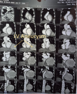
Large dilatation/outpouching arising from the left lateral wall of the left ventricle, measuring approximately 131 x 90 x 115 mm, with a neck of approximately 20 mm; abutting the left mediastinal pleura and left dome of diaphragm; suggestive of left ventricular aneurysm
| [7] | Abdelhaleem A, Leung A, Nguyen J, et al. Left ventricular aneurysm versus pseudoaneurysm: Diagnosis in the era of multi-modality imaging and 3D-printing. Echocardiography. 2023; 40: 137–142. https://doi.org/10.1111/echo.15523 |
[7]
(
Figure 2). Mild hypertrophy of the remaining left ventricle was noted. Right and left SVCs draining to the right atrium were reported. Main pulmonary artery measured about 38 mm with mild bilateral branch pulmonary artery dilatation, raising possible pulmonary hypertension.
Figure 2. CT cardiac chambers showing outpouching arising from the left lateral wall of the left ventricle with irregular hyperdensities suggestive of thrombus.
3.4. CT Coronary Angiography
Technically suboptimal study due to venous filling, poor contrast opacification, and motion blur. Left main artery was normal. LAD was type I with few proximal calcified/soft plaques causing mild 30-40% luminal narrowing. LCx showed mild 20-30% proximal stenosis with two stents in the mid and distal segments; distal contrast opacification was not clearly seen, so blocked stent could not be excluded. RCA had mild 20-30% stenosis in mid/distal segments; distal RCA/PDA/PLV were obscured. Right dominance. CAD-RADS: inconclusive study.
3.5. Chest Radiograph
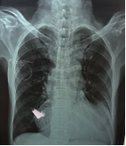
Figure 3. Chest radiograph showing left-sided cardiac contour enlargement.
Cardiomegaly/left-sided cardiac contour enlargement on radiographs (
Figure 3). No definite pleural effusion was apparent.
3.6. Laboratory Summary
Table 2. Summary of Laboratory Investigations.
Domain | Notable values |
Serology | HBsAg negative; anti-HCV negative: HIV non-reactive. |
Hematology | Mild anemia was present (hemoglobin approximately 10.9-11.2 g/dL). Platelets were within range (approximately 249-301×103/uL). WBC ranged from approximately 9.4 to 11.3×10³/uL with neutrophil predominance. ESR was 24 mm/hour. |
Coagulation | PT was reported as 18.2 sec with INR around 1.02. |
Renal/electrolytes | Creatinine ranged from approximately 0.96 to 1.24 mg/dL. Mild hyponatremia was noted (Sodium approximately 132.5-133 mmol/L). One chloride value was low (approximately 93 mmol/L). |
Biochemistry | Mild hyperbilirubinemia was recorded (total bilirubin approximately 1.8 mg/dL). LDH was elevated (approximately 537 IU/L). Total protein was low (approximately 4.89 g/dL). Random glucose was 86 mg/dL on one report and 139 mg/dL on another. |
Lipid profile | Total cholesterol approximately 90 mg/dL, LDL approximately 41 mg/dL, HDL approximately 27.9 mg/dL, triglycerides approximately 116.5 mg/dL. |
4. Differential Diagnosis
True post-infarction left ventricular aneurysm: supported by remote myocardial infarction, chronic course, ventricular outpouching, regional wall motion abnormality, and operative repair anatomy.
Left ventricular pseudoaneurysm: important alternative diagnosis because the lesion is lateral/posterolateral and has a relatively narrow neck; definitive differentiation depends on imaging, intra-operative findings, and histopathology of the aneurysmal wall.
Pericardial cyst or mediastinal mass: less likely because imaging demonstrated communication with the left ventricular cavity and associated post-infarction regional wall abnormality.
Congenital left ventricular diverticulum/aneurysm: less likely in the setting of prior myocardial infarction and LCx stenting, but can be considered if operative histology does not show post-infarction scar.
5. Operative Notes
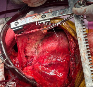
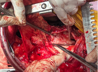
The patient was prepared for surgery under standard cardiac anesthesia and positioned in supine position. median sternotomy done. Cardio-pulmonary bypass was instituted by ascending Aorta and right atrial appendage cannulation. Del-nido cardioplegia solution was used to arrest the heart. A giant LV lateral wall aneurysm was found after extensive dissection of pericardial adhesions (
Figure 4).
Figure 4. Intra-operative picture showing giant LV aneurysm attached to lateral wall of LV after removing pericardial adhesions.
Longitudinal incision of aneurysm (
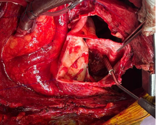
Figure 5) and removal of thrombus (
Figure 6) were performed. The aneurysm was connected to lateral wall of LV through a neck approximately 3×5 cm.
Figure 5. Intra-operative picture of incision of aneurysm.
Figure 6. Open aneurysm with thrombus present.
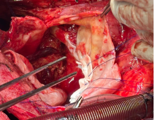
Figure 7. Closure of aneurysm neck with Teflon felt strips and polypropylene sutures.
Two strips of Teflon felt taken and placed around both the sides of oval shaped neck, five polypropylene 4-0 sutures taken in mattress fashion involving both the felt strips and neck margins (
Figure 7). Second layer taken with polypropylene 3-0 running sutures. Pericardial patch of same size taken and sewn around the closed neck with polypropylene 4-0 running sutures. there was no bleeding after surgical reconstruction. Smoothly taken patient off Cardio-pulmonary bypass. Total cross-clamp time was 47minutes. Total bypass time was 85minutes. Intra-aortic balloon pump (IABP) was placed before closure. Total 3 units of blood (PRBC) transfused intra-operatively.
6. Postoperative Course and Outcome
The patient was shifted to ICU after completion of surgery with IABP and heavy ionotropic supports (dobutamine, epinephrine and nor-epinephrine) and remained vitally stable throughout the intubation period. He was extubated twenty hours after surgery and remained stable after extubation and slowly ionotropic supports were tapered down. Postoperative echocardiographic LVEF was 35%, no residual aneurysm and mitral/tricuspid/aortic valve working normal. ICU stay of patient was 4 days post operative period and then patient shifted to ward for 1 day and discharged with stable vitals on oral antibiotics, pain killers, tablet aspirin 75mg, tablet clopidogrel 75mg, tablet rosuvastatin 10mg, tablet nitroglycerine 2.6mg and tablet torsemide 10mg. At 15 days follow-up, patient remained asymptomatic with satisfactory wound healing.
7. Discussion
7.1. Pathogenesis and Clinical Importance
Post-infarction left ventricular aneurysm develops when a transmural infarcted segment undergoes thinning, fibrosis, expansion, and paradoxical systolic bulging
| [1] | Sattar Y, Alraies MC. Ventricular Aneurysm. In: StatPearls [Internet]. Treasure Island (FL): StatPearls Publishing; updated 2023 Apr 3. |
[1]
. The resulting dyskinetic segment increases wall stress and may reduce effective stroke volume. In large aneurysms, this can produce heart failure symptoms, recurrent ischemic symptoms, mitral regurgitation due to altered geometry, ventricular arrhythmias from scar border zones, and stasis-related mural thrombus with embolic risk
| [2] | Lorusso R, Matteucci M, Lerakis S, Ronco D, Menicanti L, Sharma SK, Moreno PR. Postmyocardial Infarction Ventricular Aneurysm: JACC Focus Seminar 5/5. J Am Coll Cardiol. 2024; 83(19): 1917-1935.
https://doi.org/10.1016/j.jacc.2024.02.044 |
| [3] | Jia JH, Fu W, Sun YP, et al. Advances in Surgical Treatments of Left Ventricular Aneurysms. Rev Cardiovasc Med. 2024; 25(8): 290. https://doi.org/10.31083/j.rcm2508290 |
| [8] | Kwok CS, Bennett S, Borovac JA, et al. Predictors of left ventricular thrombus after acute myocardial infarction: a systematic review and meta-analysis. Coron Artery Dis. 2023; 34: 250–259. https://doi.org/10.1097/MCA.0000000000001223 |
[2, 3, 8]
.
This patient had a remote myocardial infarction treated with LCx stenting and later developed a giant aneurysmal outpouching from the lateral/posterolateral left ventricle. A lateral location is clinically important because LCx-related infarction can involve the lateral or posterolateral wall, and because posterior/lateral outpouchings overlap radiologically with pseudoaneurysm, especially when the neck is relatively narrow.
7.2. Role of Imaging
Multimodality imaging is central to diagnosis and surgical planning
| [2] | Lorusso R, Matteucci M, Lerakis S, Ronco D, Menicanti L, Sharma SK, Moreno PR. Postmyocardial Infarction Ventricular Aneurysm: JACC Focus Seminar 5/5. J Am Coll Cardiol. 2024; 83(19): 1917-1935.
https://doi.org/10.1016/j.jacc.2024.02.044 |
| [7] | Abdelhaleem A, Leung A, Nguyen J, et al. Left ventricular aneurysm versus pseudoaneurysm: Diagnosis in the era of multi-modality imaging and 3D-printing. Echocardiography. 2023; 40: 137–142. https://doi.org/10.1111/echo.15523 |
[2, 7]
. Echocardiography can assess ventricular function, regional wall motion, valve regurgitation, pericardial effusion, thrombus, and flow across the aneurysmal neck. CT provides excellent anatomic definition of aneurysm size, neck, relation to pleura/diaphragm, coronary stents, extracardiac thoracic findings, and vascular anomalies such as persistent left SVC. Cardiac MRI, if available and not contraindicated, is valuable for scar characterization, thrombus detection, and differentiation of true aneurysm from pseudoaneurysm
| [7] | Abdelhaleem A, Leung A, Nguyen J, et al. Left ventricular aneurysm versus pseudoaneurysm: Diagnosis in the era of multi-modality imaging and 3D-printing. Echocardiography. 2023; 40: 137–142. https://doi.org/10.1111/echo.15523 |
[7]
.
In the present case, echocardiography and CT were concordant in identifying a giant LV outpouching. CT coronary angiography, however, was inconclusive because of poor contrast and motion blur, leaving uncertainty regarding distal LCx stent patency and distal RCA/PDA/PLV visualization.
7.3. True Aneurysm Versus Pseudoaneurysm
A true aneurysm contains scarred myocardium/fibrous ventricular wall and usually has a lower risk of rupture once mature. A pseudoaneurysm is a contained rupture of the left ventricular free wall bounded by pericardium or adherent scar; it is more prone to rupture and generally warrants early surgical consideration. Features suggesting pseudoaneurysm include a narrow neck relative to sac size, abrupt discontinuity of myocardium, posterior/inferior/lateral location, and a history of recent infarction or cardiac surgery. Although histopathological examination of aneurysmal wall was not available till the time of discharge, the chronic clinical course, multimodality imaging findings and intraoperative appearance favoured a chronic post-infarctiom true left ventricular aneurysm over pseudoaneurysm
| [6] | Torchio F, Garatti A, Ronco D, Matteucci M, Massimi G, Lorusso R. Left ventricular pseudoaneurysm: the niche of post-infarction mechanical complications. Ann Cardiothorac Surg. 2022; 11(3): 290-298.
https://doi.org/10.21037/acs-2022-ami-25 |
| [7] | Abdelhaleem A, Leung A, Nguyen J, et al. Left ventricular aneurysm versus pseudoaneurysm: Diagnosis in the era of multi-modality imaging and 3D-printing. Echocardiography. 2023; 40: 137–142. https://doi.org/10.1111/echo.15523 |
| [9] | Vukovic P, et al. (2024). Surgical treatment of a left ventricular pseudoaneurysm with an extracellular matrix patch. Indian Journal of Thoracic and Cardiovascular Surgery. 2024.
https://doi.org/10.1007/s12055-023-01669-3 |
| [10] | Yeo TC, Malouf JF, Oh JK, Seward JB. Clinical profile and outcome in 52 patients with cardiac pseudoaneurysm. Annals of Internal Medicine. 1998; 128(4): 299–305. |
[6, 7, 9, 10]
.
7.4. Indications for Surgery and Operative Strategy
Management of LVA is individualized. Small, asymptomatic aneurysms may be observed with optimized medical therapy
| [2] | Lorusso R, Matteucci M, Lerakis S, Ronco D, Menicanti L, Sharma SK, Moreno PR. Postmyocardial Infarction Ventricular Aneurysm: JACC Focus Seminar 5/5. J Am Coll Cardiol. 2024; 83(19): 1917-1935.
https://doi.org/10.1016/j.jacc.2024.02.044 |
| [3] | Jia JH, Fu W, Sun YP, et al. Advances in Surgical Treatments of Left Ventricular Aneurysms. Rev Cardiovasc Med. 2024; 25(8): 290. https://doi.org/10.31083/j.rcm2508290 |
[2, 3]
. Surgical repair is usually considered when there is large aneurysm size, progressive expansion, heart failure, angina or ischemia, ventricular arrhythmia, systemic embolism or thrombus, significant valve dysfunction, pseudoaneurysm, or diagnostic/rupture concern. The operative goal is not only to remove or exclude the aneurysmal segment but also to restore a more physiological left ventricular shape and volume
| [11] | Delgado Lopez M, et al. (2024). Surgical ventricular reconstruction and intraoperative cryoablation in a patient with drug-refractory ventricular tachycardia and left ventricular thrombus: a case report. European Heart Journal – Case Reports. 2024.
https://doi.org/10.1093/ehjcr/ytae113 |
[11]
.
Surgical techniques include linear aneurysmorrhaphy, aneurysmectomy, endoventricular circular patch plasty/Dor-type repair, and other ventricular reconstruction techniques, with or without concomitant coronary artery bypass grafting or valve repair. Patch reconstruction is particularly useful in large aneurysms because it excludes non-contractile scar while preserving a more elliptical ventricular cavity and avoiding excessive reduction of LV volume
| [3] | Jia JH, Fu W, Sun YP, et al. Advances in Surgical Treatments of Left Ventricular Aneurysms. Rev Cardiovasc Med. 2024; 25(8): 290. https://doi.org/10.31083/j.rcm2508290 |
| [12] | Ruzza A, Czer LSC, Arabia F, Vespignani R, Esmailian F, Cheng W, De Robertis MA, Trento A. Left Ventricular Reconstruction for Postinfarction Left Ventricular Aneurysm: Review of Surgical Techniques. Tex Heart Inst J. 2017; 44(5): 326-335. |
| [13] | Song Y, Xu F, Feng W. Surgical treatment of left ventricular aneurysm in patients with prior myocardial infarction in the absence of obstructive coronary artery disease (MINOCA): a cohort study. J Cardiothorac Surg. 2023; 18: 304.
https://doi.org/10.1186/s13019-023-02385-0 |
| [14] | Liu Y, Cai Z, Xu L, et al. (2023). Concomitant valve surgery is associated with worse outcomes in surgical treatments of post-infarction ventricular aneurysm. Frontiers in Cardiovascular Medicine. 2023; 10: 1194374.
https://doi.org/10.3389/fcvm.2023.1194374 |
[3, 12-14]
.
7.5. Antithrombotic and Secondary Prevention Considerations
Patients with large LV aneurysms are at risk of mural thrombus because of blood stasis
| [8] | Kwok CS, Bennett S, Borovac JA, et al. Predictors of left ventricular thrombus after acute myocardial infarction: a systematic review and meta-analysis. Coron Artery Dis. 2023; 34: 250–259. https://doi.org/10.1097/MCA.0000000000001223 |
| [15] | Levine GN, McEvoy JW, Fang JC, Ibeh C, McCarthy CP, Misra A, Shah ZI, Shenoy C, Spinler SA, Vallurupalli S, Lip GYH. Management of Patients at Risk for and With Left Ventricular Thrombus: A Scientific Statement From the American Heart Association. Circulation. 2022; 146(15): e205-e223. https://doi.org/10.1161/CIR.0000000000001092 |
[8, 15]
. If thrombus is documented, anticoagulation strategy should be individualized alongside antiplatelet requirements for coronary stents and postoperative bleeding risk. Guideline-based secondary prevention after myocardial infarction and PCI should include antiplatelet therapy, high-intensity statin unless contraindicated, beta-blocker when appropriate, renin-angiotensin system inhibition/ARNI as indicated, risk-factor control, and supervised cardiac rehabilitation.
8. Unique Aspects of This Case
1) Giant size: the aneurysmal sac measured approximately 13 cm in maximum dimension on CT, making it a giant lesion with mass effect on adjacent thoracic structures.
2) Lateral/posterolateral location: this is less common than typical apical/anterior aneurysms and requires careful pseudoaneurysm differentiation.
3) Prior LCx stenting: the aneurysm location and LCx stents support an ischemic etiology involving the lateral wall.
4) Delayed presentation: presented nearly four years after myocardial infarction.
9. Conclusion
A giant post-infarction left ventricular aneurysm is a rare and potentially serious delayed complication of myocardial infarction. This case demonstrates the importance of integrating clinical history, echocardiography, CT cardiac chamber imaging, coronary assessment, and operative findings. In large lateral/posterolateral aneurysms, pseudoaneurysm must remain in the differential diagnosis until imaging and surgical inspection clarify the anatomy. The present lesion was most consistent with a giant chronic post-infarction left ventricular aneurysm based on multimodality imaging and intraoperative findings. Patch-based ventricular reconstruction under cardiopulmonary bypass is a rational surgical strategy for excluding the dyskinetic cavity and restoring left ventricular geometry
| [2] | Lorusso R, Matteucci M, Lerakis S, Ronco D, Menicanti L, Sharma SK, Moreno PR. Postmyocardial Infarction Ventricular Aneurysm: JACC Focus Seminar 5/5. J Am Coll Cardiol. 2024; 83(19): 1917-1935.
https://doi.org/10.1016/j.jacc.2024.02.044 |
| [3] | Jia JH, Fu W, Sun YP, et al. Advances in Surgical Treatments of Left Ventricular Aneurysms. Rev Cardiovasc Med. 2024; 25(8): 290. https://doi.org/10.31083/j.rcm2508290 |
| [10] | Yeo TC, Malouf JF, Oh JK, Seward JB. Clinical profile and outcome in 52 patients with cardiac pseudoaneurysm. Annals of Internal Medicine. 1998; 128(4): 299–305. |
[2, 3, 10]
.
10. Learning Points
1) Remote myocardial infarction can lead to delayed, giant left ventricular aneurysm formation, especially when infarction is transmural or reperfusion is incomplete.
2) Lateral or posterior LV outpouchings need careful differentiation between true aneurysm and pseudoaneurysm because management urgency and rupture risk differ.
3) Echocardiography defines function and valve status, while CT/MRI better define sac size, neck anatomy, relation to adjacent structures, and thrombus or scar characteristics.
4) Large or symptomatic LV aneurysms should be evaluated by a multidisciplinary heart team for surgical reconstruction, coronary management, and postoperative antithrombotic planning.
5) Surgical repair with aneurysm exclusion and left ventricular reconstruction remains an effective treatment for giant chronic post-infarction left ventricular aneurysms, restoring ventricular geometry and improving clinical outcomes.
11. Patient Consent and Ethics
This draft has been anonymized by removing patient name, identifiers, and hospital numbers. Written informed consent was obtained from the patient for publication of this case report and the accompanying clinical images. Ethical approval was not required for this single-patient case report in accordance with the institutional policy.
Abbreviations
CT | Computed Tomography |
LAD | Left Anterior Descending Coronary Artery |
LCx | Left Circumflex Coronary Artery |
RCA | Right Coronary Artery |
LVA | Left Ventricular Aneurysm |
PCI | Percutaneous Coronary Intervention |
PTCA | Percutaneous Transluminal Coronary Angioplasty |
HRCT | High-Resolution Computed Tomography |
SVC | Superior Vena Cava |
PDA | Posterior Descending Artery |
PLV | Posterolateral Ventricular Branch |
CAD-RADS | Coronary Artery Disease–Reporting and Data System |
HBsAg | Hepatitis B Surface Antigen |
HCV | Hepatitis C Virus |
HIV | Human Immunodeficiency Virus |
WBC | White Blood Cell Count |
ESR | Erythrocyte Sedimentation Rate |
PT | Prothrombin Time |
INR | International Normalized Ratio |
LDH | Lactate Dehydrogenase |
LDL | Low-Density Lipoprotein |
HDL | High-Density Lipoprotein |
IABP | Intra-Aortic Balloon Pump |
PRBC | Packed Red Blood Cells |
LVEF | Left Ventricular Ejection Fraction |
ICU | Intensive Care Unit |
MRI | Magnetic Resonance Imaging |
Author Contributions
Utsav Nandwana: Conceptualization, Data curation, Formal Analysis, Visualization, Writing – original draft
Sanjeev Devgarha: Methodology, Supervision, Validation, Writing – review & editing
Anula Sisodia: Formal Analysis, Methodology, Writing – review & editing
Krishna Kumar Mawar: Formal Analysis, Writing – review & editing
Dhruv Sharma: Formal Analysis, Writing – review & editing
Swapnil Panchal: Data Curation, Investigation
Mohit Singhal: Data Curation, Investigation
Data Availability Statement
The data supporting the findings of this case report are available from the corresponding author upon reasonable request. To protect patient privacy and confidentiality, the data are not publicly available.
Conflicts of Interest
The authors declare no conflict of interest.
References
| [1] |
Sattar Y, Alraies MC. Ventricular Aneurysm. In: StatPearls [Internet]. Treasure Island (FL): StatPearls Publishing; updated 2023 Apr 3.
|
| [2] |
Lorusso R, Matteucci M, Lerakis S, Ronco D, Menicanti L, Sharma SK, Moreno PR. Postmyocardial Infarction Ventricular Aneurysm: JACC Focus Seminar 5/5. J Am Coll Cardiol. 2024; 83(19): 1917-1935.
https://doi.org/10.1016/j.jacc.2024.02.044
|
| [3] |
Jia JH, Fu W, Sun YP, et al. Advances in Surgical Treatments of Left Ventricular Aneurysms. Rev Cardiovasc Med. 2024; 25(8): 290.
https://doi.org/10.31083/j.rcm2508290
|
| [4] |
Arora GS, Gill TK. Critical insights into the diagnosis and management of post-myocardial infarction left ventricular aneurysm of the inferior wall: a case report and brief review. Arch Med Sci Atheroscler Dis. 2024.
https://doi.org/10.5114/amsad/189733
|
| [5] |
Ba'albaki HA, Clements SD. Left ventricular aneurysm: a review. Clin Cardiol. 1989; 12(1): 5-13.
|
| [6] |
Torchio F, Garatti A, Ronco D, Matteucci M, Massimi G, Lorusso R. Left ventricular pseudoaneurysm: the niche of post-infarction mechanical complications. Ann Cardiothorac Surg. 2022; 11(3): 290-298.
https://doi.org/10.21037/acs-2022-ami-25
|
| [7] |
Abdelhaleem A, Leung A, Nguyen J, et al. Left ventricular aneurysm versus pseudoaneurysm: Diagnosis in the era of multi-modality imaging and 3D-printing. Echocardiography. 2023; 40: 137–142.
https://doi.org/10.1111/echo.15523
|
| [8] |
Kwok CS, Bennett S, Borovac JA, et al. Predictors of left ventricular thrombus after acute myocardial infarction: a systematic review and meta-analysis. Coron Artery Dis. 2023; 34: 250–259.
https://doi.org/10.1097/MCA.0000000000001223
|
| [9] |
Vukovic P, et al. (2024). Surgical treatment of a left ventricular pseudoaneurysm with an extracellular matrix patch. Indian Journal of Thoracic and Cardiovascular Surgery. 2024.
https://doi.org/10.1007/s12055-023-01669-3
|
| [10] |
Yeo TC, Malouf JF, Oh JK, Seward JB. Clinical profile and outcome in 52 patients with cardiac pseudoaneurysm. Annals of Internal Medicine. 1998; 128(4): 299–305.
|
| [11] |
Delgado Lopez M, et al. (2024). Surgical ventricular reconstruction and intraoperative cryoablation in a patient with drug-refractory ventricular tachycardia and left ventricular thrombus: a case report. European Heart Journal – Case Reports. 2024.
https://doi.org/10.1093/ehjcr/ytae113
|
| [12] |
Ruzza A, Czer LSC, Arabia F, Vespignani R, Esmailian F, Cheng W, De Robertis MA, Trento A. Left Ventricular Reconstruction for Postinfarction Left Ventricular Aneurysm: Review of Surgical Techniques. Tex Heart Inst J. 2017; 44(5): 326-335.
|
| [13] |
Song Y, Xu F, Feng W. Surgical treatment of left ventricular aneurysm in patients with prior myocardial infarction in the absence of obstructive coronary artery disease (MINOCA): a cohort study. J Cardiothorac Surg. 2023; 18: 304.
https://doi.org/10.1186/s13019-023-02385-0
|
| [14] |
Liu Y, Cai Z, Xu L, et al. (2023). Concomitant valve surgery is associated with worse outcomes in surgical treatments of post-infarction ventricular aneurysm. Frontiers in Cardiovascular Medicine. 2023; 10: 1194374.
https://doi.org/10.3389/fcvm.2023.1194374
|
| [15] |
Levine GN, McEvoy JW, Fang JC, Ibeh C, McCarthy CP, Misra A, Shah ZI, Shenoy C, Spinler SA, Vallurupalli S, Lip GYH. Management of Patients at Risk for and With Left Ventricular Thrombus: A Scientific Statement From the American Heart Association. Circulation. 2022; 146(15): e205-e223.
https://doi.org/10.1161/CIR.0000000000001092
|
Cite This Article
-
APA Style
Nandwana, U., Devgarha, S., Sisodia, A., Mawar, K. K., Sharma, D., et al. (2026). Giant Post-Infarction Left Ventricular Aneurysm of the Lateral Wall. International Journal of Cardiovascular and Thoracic Surgery, 12(4), 99-106. https://doi.org/10.11648/j.ijcts.20261204.11
 Copy
|
Copy
|
 Download
ACS Style
Nandwana, U.; Devgarha, S.; Sisodia, A.; Mawar, K. K.; Sharma, D., et al. Giant Post-Infarction Left Ventricular Aneurysm of the Lateral Wall. Int. J. Cardiovasc. Thorac. Surg. 2026, 12(4), 99-106. doi: 10.11648/j.ijcts.20261204.11
Copy
|
Download
AMA Style
Nandwana U, Devgarha S, Sisodia A, Mawar KK, Sharma D, et al. Giant Post-Infarction Left Ventricular Aneurysm of the Lateral Wall. Int J Cardiovasc Thorac Surg. 2026;12(4):99-106. doi: 10.11648/j.ijcts.20261204.11
Copy
|
Download
-
@article{10.11648/j.ijcts.20261204.11,
  author = {Utsav Nandwana and Sanjeev Devgarha and Anula Sisodia and Krishna Kumar Mawar and Dhruv Sharma and Swapnil Panchal and Mohit Singhal},
  title = {Giant Post-Infarction Left Ventricular Aneurysm of the Lateral Wall},
  journal = {International Journal of Cardiovascular and Thoracic Surgery},
  volume = {12},
  number = {4},
  pages = {99-106},
  doi = {10.11648/j.ijcts.20261204.11},
  url = {https://doi.org/10.11648/j.ijcts.20261204.11},
  eprint = {https://article.sciencepublishinggroup.com/pdf/10.11648.j.ijcts.20261204.11},
  abstract = {Background: Left ventricular aneurysm is an uncommon but important late mechanical complication of transmural myocardial infarction. Giant aneurysms can cause heart failure, angina, ventricular arrhythmia, thromboembolism, and diagnostic confusion with pseudoaneurysm or mediastinal masses. Case presentation: A 58-year-old male with a history of acute myocardial infarction in 2022 treated abroad by percutaneous coronary intervention/angioplasty with two stents in the left circumflex artery was evaluated for a large left ventricular outpouching. Earlier surgical advice had reportedly been declined, and the patient was managed medically until re-presentation in 2026. Echocardiography showed a large cystic aneurysmal lesion communicating with the posterior left ventricular wall through an approximately 3 cm neck, with left ventricular ejection fraction around 40%, mild left ventricular systolic dysfunction, grade I diastolic dysfunction, mild aortic regurgitation, and mild tricuspid regurgitation with an estimated right ventricular systolic pressure of approximately 36 mmHg. CT cardiac chamber imaging demonstrated cardiomegaly and a giant outpouching arising from the left lateral wall of the left ventricle, measuring approximately 131 x 90 x 115 mm with a 20 mm neck. CT coronary angiography showed mild plaque burden in the LAD, LCx, and RCA, with uncertain distal LCx stent patency. The patient underwent open surgical repair with exclusion of the aneurysmal cavity and ventricular reconstruction with primary multilayer closure of neck using Teflon felt under cardiopulmonary bypass. Conclusion: This case highlights the value of multimodality imaging for defining aneurysm size, neck anatomy, coronary status, and associated thoracic findings. Giant post-infarction aneurysms, especially when large, symptomatic, or anatomically high risk, require individualized heart-team assessment and often surgical reconstruction to restore ventricular geometry and reduce complications.},
 year = {2026}
}
Copy
|
Download
-
TY  - JOUR
T1  - Giant Post-Infarction Left Ventricular Aneurysm of the Lateral Wall
AU  - Utsav Nandwana
AU  - Sanjeev Devgarha
AU  - Anula Sisodia
AU  - Krishna Kumar Mawar
AU  - Dhruv Sharma
AU  - Swapnil Panchal
AU  - Mohit Singhal
Y1  - 2026/07/24
PY  - 2026
N1  - https://doi.org/10.11648/j.ijcts.20261204.11
DO  - 10.11648/j.ijcts.20261204.11
T2  - International Journal of Cardiovascular and Thoracic Surgery
JF  - International Journal of Cardiovascular and Thoracic Surgery
JO  - International Journal of Cardiovascular and Thoracic Surgery
SP  - 99
EP  - 106
PB  - Science Publishing Group
SN  - 2575-4882
UR  - https://doi.org/10.11648/j.ijcts.20261204.11
AB  - Background: Left ventricular aneurysm is an uncommon but important late mechanical complication of transmural myocardial infarction. Giant aneurysms can cause heart failure, angina, ventricular arrhythmia, thromboembolism, and diagnostic confusion with pseudoaneurysm or mediastinal masses. Case presentation: A 58-year-old male with a history of acute myocardial infarction in 2022 treated abroad by percutaneous coronary intervention/angioplasty with two stents in the left circumflex artery was evaluated for a large left ventricular outpouching. Earlier surgical advice had reportedly been declined, and the patient was managed medically until re-presentation in 2026. Echocardiography showed a large cystic aneurysmal lesion communicating with the posterior left ventricular wall through an approximately 3 cm neck, with left ventricular ejection fraction around 40%, mild left ventricular systolic dysfunction, grade I diastolic dysfunction, mild aortic regurgitation, and mild tricuspid regurgitation with an estimated right ventricular systolic pressure of approximately 36 mmHg. CT cardiac chamber imaging demonstrated cardiomegaly and a giant outpouching arising from the left lateral wall of the left ventricle, measuring approximately 131 x 90 x 115 mm with a 20 mm neck. CT coronary angiography showed mild plaque burden in the LAD, LCx, and RCA, with uncertain distal LCx stent patency. The patient underwent open surgical repair with exclusion of the aneurysmal cavity and ventricular reconstruction with primary multilayer closure of neck using Teflon felt under cardiopulmonary bypass. Conclusion: This case highlights the value of multimodality imaging for defining aneurysm size, neck anatomy, coronary status, and associated thoracic findings. Giant post-infarction aneurysms, especially when large, symptomatic, or anatomically high risk, require individualized heart-team assessment and often surgical reconstruction to restore ventricular geometry and reduce complications.
VL  - 12
IS  - 4
ER  -
Copy
|
Download

 ,
Sanjeev Devgarha
,
Sanjeev Devgarha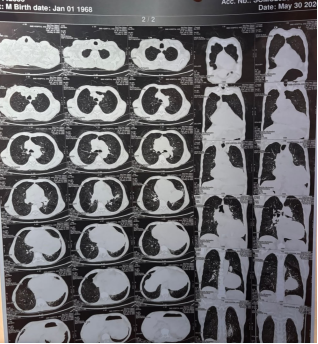



 Share This Article
 Twitter
 Linked In
 Facebook